



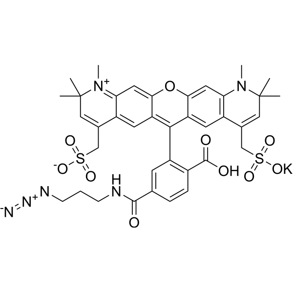
AF 594 azide
AF 594 azide is a red-emitting alkyne-azide click chemistry (CuAAC) fluorescent probe, with an Alexa Fluor 594 analog fluorophore (Ex/Em ≈ 590/617 nm) at one end of the molecule and an azide (-N3) reactive group at the other end. Under Cu (I) catalysis, AF 594 azide undergoes efficient covalent coupling with nucleic acids, proteins, lipids, or materials bearing terminal alkynes (-C≡CH), and is a versatile dye for fluorescent labeling of alkyne-modified metabolic markers (EdU/EdA/EU and other alkynyl nucleosides), alkynyl unnatural amino acids, and alkynyl lipids/liposomes.
For research use only. We do not sell to patients.
- Formula: C38H39KN6O10S2
- Molecular Weight:842.98
-
Storage:
Please store the product under the recommended conditions in the Certificate of Analysis.

To place orders, for customer services and technical support, please contact: MedChemExpress USA
Tel: 609-228-6898 E-mail: [email protected] [email protected]


-
 Biological Activity
-
Chemical Information
- Protocols
- Purity & Documentation
- References
-
 Help & FAQs


Biological Activity
Description
In Vitro
1. Stock Solution Preparation
1.1 Solvent: anhydrous DMSO[3].
1.2 Recommended concentration: Generally, a high-concentration stock solution of 1-10 mM is recommended, aliquoted and stored protected from light.
2. Working Solution Preparation
2.1 Diluent: PBS.
2.2 Working concentration: AF 594 azide is dissolved in freshly prepared click staining mixture containing 1 mM CuSO4, 10 mM sodium ascorbate, 1 mM Tris (3-hydroxypropyltriazolylmethyl) amine (THPTA) (HY-W021042), and 10 mM Aminoguanidine (HY-B1041A)[2].
2.3 Prepare fresh before use.
3. Staining Procedure
3.1 For liposomes bearing terminal alkyne residues[1]:
Liposomes bearing terminal alkyne residues are derivatized with AF 594 azide via copper-catalyzed azide-alkyne cycloaddition (CuAAC) in a one-pot orthogonal click coupling reaction.
3.2 For eukaryotic cells co-incubated with AF 594 azide-labeled liposomes[1]:
Eukaryotic cells are co-incubated with AF 594 azide-labeled liposomes, and rapid staining of the cell membrane is observed within 30 min; experiments show that only cholesterol-anchored liposomes integrate into the cell membrane.
3.3 For HeLa-Ohio cells infected with EdA/EdC-HAdV-C5 and permeabilized with SLO[2]:
HeLa-Ohio cells are cultured in coverslips in 24-well plates; EdA/EdC-HAdV-C5 (1 mg) is bound on ice for 60 min and internalized at 37 °C; Streptolysin O (HY-135416) binds to the plasma membrane and is briefly incubated at 37°C for permeabilization (without fixing cells); AF 594 azide is perfused at room temperature for staining for 60 min; conventionally fixed cells are stained with fresh click staining mixture at room temperature in the dark for 2 h (30 min for vaccinia virus samples); after staining, cells are washed, fixed with PFA, permeabilized with Triton X-100, and then immunostained for hexon.
3.4 For cells used for RNA detection[3]:
Cells are cultured in minimum essential medium containing glutamine, penicillin, Streptomycin (HY-B1906), and 10% FBS, seeded at 10,000-20,000 cells/well in 1 cm2 microculture plates, and grown to near confluence; RNA is pulse-labeled by co-incubation with 1 mM 5-Ethynyluridine (HY-141140) for 30 min and fixed with 4% PFA/PBS; labeled transcripts are detected using AF 594 azide (Click-iT RNA Imaging Kit).
3.5 For the αS56-140-C56 fragment containing propargyl-tyrosine[4]:
AF 594 azide is attached to the propargyl-tyrosine site via copper-catalyzed cycloaddition reaction.
3.6 For live HeLa or U2OS cells after KBrO3-induced lipid peroxidation or POVPC exposure[5]:
KBrO3 group: cells are exposed to 5 mM KBrO3 for 17 h, washed, and incubated with 10 µM FLipA-HIPS at 37°C for 4 h; POVPC group: cells are incubated with 25 µM POVPC for 1 h, and 10 µM FLipA-HIPS is directly added and incubated at 37°C for 2 h; after FLipA-HIPS staining, cells are fixed with cold methanol, and the FLipA-HIPS reaction products in fixed cells are labeled with AF 594 azide via CuAAC.
4. Controls
4.1 Negative control: tetrazine-bearing liposomes do not react with AF 594 azide[1].
4.2 Post-processing control: fluorescence correlation spectroscopy/size exclusion chromatography confirms no residual free dye azide after post-processing[1].
4.3 Untreated control: cells without KBrO3 (0 mM) or without POVPC (0 µM)[5].
4.4 ER-tracker control: after KBrO3 and FLipA-HIPS application, cells are washed and incubated with 2 µM BODIPY FL Glibenclamide as ER-tracker at 37°C for 30 minutes, followed by confocal microscopy examination[5].
4.5 MitoTracker Red control: after KBrO3 and FLipA-HIPS application, cells are washed and incubated with 2 µM BODIPY FL Glibenclamide at 37°C for 30 min, followed by confocal imaging[5].
4.6 Viability control: 2.5 µg/mL Propidium Iodide (HY-D0815) is added for 5 minutes before confocal microscopy examination[5].
4.7 Unclicked/uninfected control: cells or virus without metabolic labeling (no alkynyl nucleoside) are incubated according to the same click procedure to subtract background and set fluorescence cutoff values[2][3].
5. Detection and Analysis
5.1 Instrument types[1][2][4][5]: confocal laser scanning microscopy; gated STED super-resolution microscopy; deconvolution or confocal laser immunofluorescence microscopy; confocal microscopy; single-molecule FRET (smFRET), fluorescence correlation spectroscopy, and size exclusion chromatography with fluorescence quantification.
5.2 Wavelength and filter settings[4][5]: AF 594 azide is excited at 561 nm. For FLipA-HIPS, the excitation wavelength is 405 nm; for ER-tracker, the excitation wavelength is 488 nm. For smFRET, 485 nm and 560 nm pulsed lasers are adjusted to 30 μW at 40 MHz before entering the microscope. An HQ585LP dichroic mirror combined with ET525/50M and HQ600LP filters is used to separate excitation and emission, and signals are detected by photodiodes.
5.3 Result analysis:
5.3.1 AF 594 azide-labeled cholesterol-anchored liposomes exhibit red fluorescence in dual-labeling experiments and rapidly stain the RBE4 cell membrane within 30 min[1].
5.3.2 At 30 minutes post-infection, AF 594 azide spots colocalize with anti-hexon spots in the cytoplasm and near the nuclear membrane. At 90 minutes post-infection, strong punctate signals of free vDNA appear in the cytoplasm and nucleus; co-staining with anti-protein VII antibody at 150 min shows that protein VII localizes in the nucleus[2].
5.3.3 After EU pulse, nascent RNA shows strong red staining plaques with weak background[3].
MedChemExpress (MCE) has not independently confirmed the accuracy of these methods. They are for reference only.


Chemical Information
-
Molecular Weight 842.98
-
Formula C38H39KN6O10S2
-
SMILES
CN1C(C)(C=C(C2=C1C=C(C3=C2)OC4=CC5=[N+](C(C)(C=C(C5=CC4=C3C6=C(C=CC(C(NCCCN=[N+]=[N-])=O)=C6)C(O)=O)CS(=O)([O-])=O)C)C)CS(=O)(O[K])=O)C
-
Shipping
Room temperature in continental US; may vary elsewhere.
-
Storage
Please store the product under the recommended conditions in the Certificate of Analysis.


Purity & Documentation
References

Calculators
Concentration (start) × Volume (start) = Concentration (final) × Volume (final)


AF 594 azide Related Classifications
HY-D2161 Related Classifications
 Powered by Bioz