



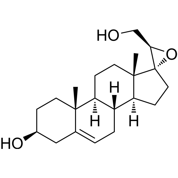
BNN27

Based on 1 Customer Validation
BNN27 is a blood-brain barrier-penetrant, selective NGF receptor (TrkA and p75NTR) activator. BNN27 activates ERK1/2/AKT. BNN27 enhances NGF-induced differentiation/axonal growth and inhibits JNK after NGF withdrawal. BNN27 reduces Amyloid-β plaques, gliosis, and pro-inflammatory cytokines, and rescues synaptic/cholinergic deficits, neurogenesis, and working memory. BNN27 can be used in research on Alzheimer's disease, amyotrophic lateral sclerosis, diabetic retinopathy, inflammatory hyperalgesia, anxiety disorders, and multiple sclerosis.
For research use only. We do not sell to patients.
- Purity : 99.54%
- CAS No.: 1195795-81-3
- Formula: C21H32O3
- Molecular Weight:332.48
-
Storage:
4°C, protect from light
* In solvent : -80°C, 6 months; -20°C, 1 month (protect from light)

To place orders, for customer services and technical support, please contact: MedChemExpress USA
Tel: 609-228-6898 E-mail: [email protected] [email protected]


-
 Biological Activity
-
Chemical Information
-
Solvent & Solubility
- Protocols
- Purity & Documentation
- References
-
 Help & FAQs


Biological Activity
Description
IC50 & Target
[1]|
TrkA |
ERK1 |
ERK2 |
In Vitro
BNN27 (18 h) shows high passive BBB permeability in the BBB-PAMPA assay, with a Pe of 12.7 μcm/s[2].
BNN27 (10 μM; 2 h) is not a BCRP substrate in Caco-2 monolayer cells, with an efflux ratio of 1.5[2].
BNN27 (1 pM-1 μM) effectively displaces [3H]-DHEA bound to membranes of HEK293TrkA transfected cells (Ki = 1.8 nM)[3].
Immobilized BNN27 (4°C overnight) directly pulls down recombinant TrkA[3] in vitro.
BNN27 (100 nM; 15 min) activates TrkA by inducing phosphorylation of Y490, Y674/675, and Y785 in CHOTrkA cells, but does not activate TrkB or TrkC[3].
BNN27 (100 nM; 60 min) promotes TrkA localization to lipid raft microdomains in HEK293TrkA cells[3].
MedChemExpress (MCE) has not independently confirmed the accuracy of these methods. They are for reference only.
-
Cell Line:CHO cells transfected with TrkA, TrkB, or TrkC cDNA
-
Concentration:100 nM (BNN27)
-
Incubation Time:15 min
-
Result:Induced phosphorylation of TrkA at Y490, Y674/675, and Y785 in CHOTrkA transfectants.
However, BNN27 did not induce Y490 phosphorylation in CHO cells transfected with TrkB or TrkC cDNA.
-
Cell Line:PC12 cells transfected with shRNAs against TrkA
-
Concentration:100 nM (BNN27)
-
Incubation Time:24 h
-
Result:FACS analysis of cells stained for cleaved Caspase-3 revealed that exposure to BNN27 reduced the number of apoptotic cells following serum deprivation from 34% (controls) to 15.33% (n=4).
Suppression of TrkA expression by shRNA almost completely reversed the anti-apoptotic effect of BNN27.
-
Cell Line:HEK293 cells transfected with TrkA plasmid
-
Concentration:100 nM (BNN27)
-
Incubation Time:60 min
-
Result:Exposure for 60 min to BNN27 caused the segregation of TrkA into lipid raft fractions identified by the presence of Flotillin-1.
In Vivo
BNN27 (10 mg/kg; intraperitoneal injection; once every two days; 4 weeks) reverses diabetes-induced loss of bNOS/TH amacrine cell markers and reduces cleaved caspase-3, but does not reduce TUNEL+ retinal cell death in STZ diabetic rats[6].
BNN27 (2.86 mg/day; subcutaneous implantation; 21-day release; implanted on day 8 and day 29) protects mature oligodendrocytes and myelin during Cuprizone-induced demyelination while reducing microgliosis and astrogliosis[9].
BNN27 (2.86 mg/day; subcutaneous implantation; 21-day release; during 3-week remyelination after 6 weeks of Cuprizone treatment) does not improve remyelination after Cuprizone (HY-W115718) withdrawal, but reduces OPC number and glial cell activation[9].
BNN27 (3-6 mg/kg; intraperitoneal injection; single injection 40 minutes before testing) increases the time spent in the light compartment of the light-dark box, indicating an anxiolytic-like effect in the light-dark box test[8].
MedChemExpress (MCE) has not independently confirmed the accuracy of these methods. They are for reference only.
-
Animal Model:C57Bl/6J background (male 5xFAD transgenic and age-matched male wild-type C57Bl/6J littermates; 1.5 months old at start; average weight ≈30 g)[1]
-
Dosage:10 mg/kg/day over 60 days (18 mg total; average animal weight ≈30 g)
-
Administration:subcutaneous; continuous release; 60 days
-
Result:Hippocampal Aβ-immunoreactive plaque load (6E10): 5xFAD mice receiving BNN27 had 13 plaques vs 5xFAD placebo mice with 23.78 plaques (n = 9).
Hippocampal GFAP-positive area: 5xFAD mice receiving BNN27 had 6.8585% vs 5xFAD placebo mice with 14.43% (n = 7-9).
Hippocampal IBA1-positive area: 5xFAD mice receiving BNN27 had 5.512% vs 5xFAD placebo mice with 11.75% (n = 7-9).
CA2 mossy fiber Synaptophysin-positive area: 5xFAD mice receiving BNN27 had 46.34% vs 5xFAD placebo mice with 16.68% (n = 6-9).
CA2 mossy fiber Synapsin I-positive signal: 5xFAD mice receiving BNN27 had 44.01% vs 5xFAD placebo mice with 21.79% (n = 6-9).
Basal forebrain ChAT+ soma size: 5xFAD mice showed reduced ChAT+ soma size vs WT (52.00 vs 67.9832); BNN27 administration restored the size of cholinergic neurons in 5xFAD mice.
Basal forebrain p75NTR+/ChAT+ neurons: number was increased in 5xFAD vs WT; BNN27 administration reduced the number of p75NTR+/ChAT+ neurons in 5xFAD mice compared with 5xFAD placebo.
Basal forebrain TrkA+/ChAT+ neurons: no change was observed in the number of TrkA+/ChAT+ neurons.
Entorhinal cortex neurofilament: BNN27 did not restore the genotype-observed reduction in neurofilament levels.
Entorhinal cortex myelin: no effect of BNN27 administration on the myelin condition.
T-maze spontaneous alternation performance: 5xFAD mice receiving BNN27 had 0.643 vs 5xFAD placebo mice with 0.4625.
Dentate gyrus BrdU+/NeuN+ neurons: 5xFAD mice receiving BNN27 had 858 vs 5xFAD placebo mice with 456.
Dentate gyrus DCX+ neurons: 5xFAD mice receiving BNN27 had 106.8 vs 5xFAD placebo mice with 82.2.
Proteomic rescue: BNN27 administration rescued 16 out of 47 down-regulated proteins by more than 10% and 38 out of 81 up-regulated proteins by more than 10%.
BNN27 administration rescued 54 out of 128 dys-regulated proteins.
In the 5xFAD+BNN27/WT comparison, 277 proteins were dys-regulated; 149 were regulated only in this comparison and 128 overlapped with the 5xFAD/WT comparison (5xFAD/WT comparison alone: 81 proteins dys-regulated).
The most rescued protein was Sptbn1; Ncstn showed minor rescue (−6%).
-
Animal Model:C57BL/6 (ngf+/-; pregnant females; embryos at E13.5/E14)[3]
-
Dosage:3 mg/day
-
Administration:i.p.; daily; gestation day 3 to E13.5
-
Result:Significantly reduced the number of Caspase-3 positive neurons in the DRG to levels comparable to ngf+/+ embryos; reported as 31 vs 19 Caspase-3 positive neurons per section (n=6).
-
Animal Model:Sprague-Dawley (adult female and male, 180-300 g)[4]
-
Dosage:19 mM; 94 mM; 470 mM
-
Administration:topical ocular (eye drops); daily for 7 days beginning 4 weeks post-STZ
-
Result:NFL-IR was 84.7% of mean gray area/section at 94 mM, 87.2% at 470 mM, and 62.5% at 19 mM (no effect).
bNOS-IR was 88.3% of cells/section at 94 mM, 93.0% at 470 mM, and 49.2% at 19 mM (no effect).
TH-IR was 91.6% of cells/section at 94 mM, 75.0% at 470 mM, and 25.0% at 19 mM (no effect).
TrkA phosphorylation: BNN27 94 mM produced a 1.8-fold increase in phospho/total TrkA ratio compared with non-treated diabetic retinas.
ERK1/2 phosphorylation: BNN27 94 mM produced 2.00 phospho/total ERK1/2 and approximately 2-fold increase compared with control retinas.
Co-administration of the specific TrkAR inhibitor (10^-3 M, intravitreal) reversed the BNN27-dependent effects at the optimum BNN27 dose of 94 mM on NFL-, bNOS-, and TH-IR compared with diabetic retinas treated with BNN27 alone; no statistically significant differences were observed in diabetic non-treated rat retinas in the presence or absence of the TrkAR inhibitor.
-
Animal Model:Wistar (male and female; 3-month-old; outbred; non-husbanded; normally cycling females; group-housed 3-4 per cage)[5]
-
Dosage:10 mg/kg; 30 mg/kg; 90 mg/kg (open field test only)
-
Administration:i.p.; single injection 30 min before each behavioral test; 90 mg/kg only before open field test
-
Result:In the open field test, BNN27 90 mg/kg reduced horizontal distance travelled and vertical rearing counts in both sexes.
10 mg/kg and 30 mg/kg did not differ from vehicle.
In the light/dark box test, BNN27 30 mg/kg reduced the total number of transitions between the light and dark compartments.
Time spent in the illuminated compartment was not affected by BNN27.
In the hole-board test, BNN27 10 mg/kg and 30 mg/kg reduced the total number of holes explored in males only.
Both doses also increased the latency to explore the first hole and reduced the hole-board working memory index in males only.
No BNN27 effects were observed in female rats in the hole-board test.
-
Animal Model:Wistar (male and female; 3-month-old; outbred; non-husbanded; normally cycling females; group-housed 3-4 per cage)[5]
-
Dosage:10 mg/kg; 30 mg/kg
-
Administration:i.p.; three injections at 23, 5, and 1 h before forced swim test session
-
Result:Did not affect immobility, swimming, or climbing duration during the forced swim test session.
Head-shake behavior was also not affected by BNN27.
-
Animal Model:Wistar (male and female; 3-month-old; outbred; non-husbanded; normally cycling females; group-housed 3-4 per cage)[5]
-
Dosage:10 mg/kg; 30 mg/kg
-
Administration:i.p.; single injection 30 min before decapitation and tissue/blood sampling
-
Result:In the hippocampus, BNN27 10 mg/kg increased GABA and glutamine levels in both sexes.
Hippocampal glutamate levels were not affected by BNN27.
10 mg/kg increased the hippocampal glutamine/glutamate ratio, indicating increased glutamate turnover, in both sexes.
In the prefrontal cortex, BNN27 10 mg/kg increased glutamate levels in males only.
Prefrontal GABA, glutamine, and glutamate turnover were not affected by BNN27.
30 mg/kg decreased testosterone serum levels in males only, whereas BNN27 10 mg/kg did not.
30 mg/kg increased progesterone serum levels in both sexes; BNN27 10 mg/kg did not.
Did not affect estrogen serum levels.
-
Animal Model:Sprague-Dawley (male and female, 180-300 g)[6]
-
Dosage:2 mg/kg i.p. daily for 7 days; 10 mg/kg i.p. daily for 7 days; 50 mg/kg i.p. daily for 7 days; 10 mg/kg i.p. daily for 7 days with intravitreal TrkAR inhibitor (10^-3 mol/L) on days 1, 4, and 6
-
Administration:i.p.; daily for 7 days; intravitreal TrkAR inhibitor on days 1, 4, and 6
-
Result:At 10 and 50 mg/kg reversed the diabetes-induced reduction in NFL-IR, bNOS-IR, and TH-IR; 2 mg/kg had no effect.
At 10 and 50 mg/kg increased TrkA Y490 phosphorylation to similar levels; 2 mg/kg had no effect.
Co-administered intravitreal TrkAR inhibitor reversed the BNN27 (10 mg/kg)-induced restoration of NFL-IR, bNOS-IR, and TH-IR compared with diabetic retinas treated with BNN27 alone.
Co-administered intravitreal TrkAR inhibitor reversed the BNN27 (10 mg/kg)-dependent increase in TrkA Y490 phosphorylation.
At 10 and 50 mg/kg attenuated the diabetes-induced increase in p75NTR-IR; 10 mg/kg reversed p75NTR expression.
Had no effect on the total number of TUNEL+ cells in the diabetic retina.
In retinas of diabetic rats treated with BNN27 (10 mg/kg), TUNEL+ cells and bNOS immunoreactivity were not colocalized.
Decreased cleaved caspase-3 immunoreactivity.
10 mg/kg: GFAP-IR 111.0% of mean gray area; 50 mg/kg: GFAP-IR 109.5%.
Diabetic retinas showed 149.0% and control retinas 100.0%.
10 mg/kg: Iba-1-positive cells 67 per mm2; 50 mg/kg: Iba-1-positive cells 113 per mm2.
Diabetic retinas showed 255 per mm2 and control retinas 100 per mm2.
10 mg/kg decreased TNFα and increased IL-10 and IL-4; 50 mg/kg reduced IL-1β.
-
Animal Model:Sprague-Dawley (male and female, 180-300 g)[6]
-
Dosage:10 mg/kg i.p. every second day for 4 weeks
-
Administration:i.p.; every second day; 4 weeks
-
Result:Did not reduce the number of TUNEL+ cells per mm2 compared with the diabetic retina.
Reversed the diabetes-induced attenuation of bNOS and TH immunoreactive cells.
Decreased cleaved caspase-3 immunoreactivity.
-
Animal Model:C57BL6x1291Sv (adult male, 8-12 weeks)[7]
-
Dosage:100 mg/kg
-
Administration:i.p.; once daily; for 4 consecutive days
-
Result:Reversed CFA-induced inflammatory hyperalgesia, with increased paw withdrawal latency at 3, 6, and 24 h following CFA.
At 6 h, increased leukocyte infiltration and MPO activity in the inflamed paw.
At 24 h, decreased immune cell infiltration and MPO activity compared with controls.
No effect on paw edema.
At 6 h, increased IL-6, TNF-α, and IL-10 levels in the inflamed paw and plasma.
At 24 h, decreased TNF-α in the inflamed paw and plasma.
IL-6 unchanged in the inflamed paw but decreased in plasma.
IL-10 remained elevated in the inflamed paw and plasma.
IL-1β in the inflamed paw showed no differences between groups at 24 h.
Plasma corticosterone was not different at 6 h, but was lower at 24 h.
Increased iNOS mRNA in the inflamed paw at 6 and 24 h.
Increased NGF mRNA in the inflamed paw at 6 h.
No change at 24 h.
At 6 h, total AKT2 unchanged, p-AKT2 and p-AKT2/total AKT2 ratio reduced.
At 24 h, total AKT2 and p-AKT2 increased, but p-AKT2/total AKT2 ratio unchanged.
No changes in AKT1.
Increased μ-opioid receptor mRNA at 6 h.
Increased POMC mRNA at 6 h and decreased POMC mRNA at 24 h.
Increased PENK mRNA at 24 h.
Decreased NGF mRNA in DRG at 6 and 24 h.
Decreased TrkA mRNA in the inflamed paw and DRG at 6 h.
At 24 h increased TrkA mRNA in DRG but no change in the inflamed paw.
No changes in p75NTR or substance P.
Increased serum β-endorphin.
-
Animal Model:C57BL6x1291Sv (adult male, 8-12 weeks)[7]
-
Dosage:10 mg/kg
-
Administration:i.p.; once daily; for 4 consecutive days
-
Result:Affected paw withdrawal latency only at 3 h following the onset of inflammation.
No effect on leukocyte infiltration at 3 h.
Decreased inflamed paw TNF-α.
No effect on IL-6 and IL-10 in the inflamed paw or plasma.
Increased μ-opioid receptor, POMC, and PENK mRNA at 3 h.
Decreased NGF mRNA in DRG at 3 h.
No changes in TrkA mRNA in the inflamed paw or DRG at 3 h.
-
Animal Model:C57BL6x1291Sv (adult male, 8-12 weeks)[7]
-
Dosage:100 mg/kg
-
Administration:i.p.; single dose 30 min before CFA
-
Result:Effective only 3 h following the onset of inflammation.
Did not alter leukocytic infiltration at 3 h.
Did not affect cytokine protein levels in the inflamed paw and plasma.
Decreased plasma corticosterone at 24 h.
Increased POMC mRNA at 3 h.
Decreased NGF mRNA in DRG at 3 h.
Increased TrkA mRNA in the inflamed paw and decreased TrkA mRNA in DRG.
-
Animal Model:C57BL6x1291Sv (adult male, 8-12 weeks)[7]
-
Dosage:10 mg/kg
-
Administration:i.p.; single dose 30 min before CFA
-
Result:Had no effect on paw withdrawal latency.
-
Animal Model:C57BL6x1291Sv (adult male, 8-12 weeks)[7]
-
Dosage:100 mg/kg and 10 mg/kg
-
Administration:i.p.; single dose 30 min after CFA
-
Result:Did not produce any effect on paw withdrawal latency for either dose.
-
Animal Model:C57BL6x1291Sv (adult male, 8-12 weeks)[7]
-
Dosage:100 mg/kg (BNN27); 20 µL (10-5 M) (TrkA inhibitor)
-
Administration:i.p.; once daily; for 4 consecutive days (BNN27); intraplantar; once daily; for 4 consecutive days (TrkA inhibitor, simultaneously with BNN27)
-
Result:Co-administration of BNN27 with TrkA inhibitor did not significantly alter paw withdrawal latency compared with controls at any time point tested.
Co-administration significantly decreased TNF-α and IL-10 at 24 h from the induction of inflammation, while IL-6 remained unchanged.
NGF mRNA levels in the inflamed paw were significantly elevated in mice co-receiving the TrkA inhibitor, whereas in DRG NGF mRNA levels remained significantly lower following TrkA inhibition.
No significant changes were observed in the mRNA levels of opioid peptide precursors and receptors after co-administration of BNN27 with the TrkA inhibitor.
-
Animal Model:C57BL6x1291Sv (adult male, 8-12 weeks)[7]
-
Dosage:100 mg/kg (BNN27); 5 mg/kg (20 µL) (naloxone-HCl)
-
Administration:i.p.; once daily; for 4 consecutive days (BNN27); intraplantar; single dose 30 min before CFA (naloxone-HCl)
-
Result:Naloxone injection dramatically reduced paw withdrawal latency in mice receiving BNN27 compared with those receiving BNN27 only.
Co-administration of BNN27 and naloxone significantly reduced β-endorphin protein levels and PENK mRNA levels in the inflamed paw compared with mice receiving BNN27 only at 24 h from the onset of inflammation.
-
Animal Model:Wistar (male, 3 months old, 250-300 g)[8]
-
Dosage:1 mg/kg, 3 mg/kg, and 6 mg/kg
-
Administration:i.p.; single injection 40 min before testing
-
Result:Administration of BNN27 did not affect the first entry into the dark chamber or the number of transitions between the two compartments.
Interestingly, BNN27 significantly increased the total time spent in the light chamber.
Vehicle-treated rats spent significantly less time in the lit chamber compared with rats treated with 3 or 6 mg/kg BNN27.
1 mg/kg did not increase time in the light chamber.\nTime spent in the central zone: BNN27 1 mg/kg, 9 s; BNN27 3 mg/kg, 14.5 s; BNN27 6 mg/kg, 14.3 s.
Number of squares crossed: BNN27 1 mg/kg, 98.4; BNN27 3 mg/kg, 94; BNN27 6 mg/kg, 99.
Number of rearings: BNN27 1 mg/kg, 32.5; BNN27 3 mg/kg, 35.6; BNN27 6 mg/kg, 35.3.
Grooming duration: BNN27 1 mg/kg, 6.8 s; BNN27 3 mg/kg, 7.3 s; BNN27 6 mg/kg, 4.9 s.
Did not affect the number of squares crossed or rearing episodes.
Appeared to reduce grooming duration, but this effect did not reach statistical significance.
Administration of BNN27 increased the time spent in the central zone of the apparatus.
Rats treated with 3 and 6 mg/kg BNN27 spent more time in the central zone of the OF apparatus compared with their vehicle-treated cohorts.
-
Animal Model:Wistar (male, 3 months old, 250-300 g)[8]
-
Dosage:1 mg/kg, 3 mg/kg, and 6 mg/kg
-
Administration:i.p.; single injection on day 3, 40 min before testing
-
Result:Administration of BNN27 significantly reduced freezing duration.
Freezing levels of rats treated with 3 and 6 mg/kg BNN27 were significantly lower compared with those expressed by vehicle-treated animals.
1 mg/kg did not reduce freezing behavior.
-
Animal Model:Wistar (male, 3 months old, 250-300 g)[8]
-
Dosage:3 mg/kg and 6 mg/kg; 5-HT2C receptor agonist 0.6 mg/kg
-
Administration:i.p.; single injection 40 min before testing; 5-HT2C receptor agonist i.p. 10 min before testing
-
Result:Grooming duration (median): vehicle + BNN27 3 mg/kg, 111.5 s; vehicle + BNN27 6 mg/kg, 104 s; 5-HT2C receptor agonist 0.6 mg/kg + BNN27 3 mg/kg, 174.5 s; 5-HT2C receptor agonist 0.6 mg/kg + BNN27 6 mg/kg, 189.5 s.
Rats receiving 5-HT2C receptor agonist plus vehicle displayed higher self-grooming compared with all other experimental groups including 5-HT2C receptor agonist + BNN27 3 mg/kg and 5-HT2C receptor agonist + BNN27 6 mg/kg.
Acute administration of BNN27 (3 and 6 mg/kg) attenuated excessive self-grooming caused by 5-HT2C receptor agonist.
Did not affect grooming activity in control animals.
Appeared to counteract the effects of 5-HT2C receptor agonist on horizontal (hypomotility) and vertical activity (decrease of rearings), but this action did not achieve statistical significance.
-
Animal Model:C57B110/CBA (male, 8 weeks old)[9]
-
Dosage:2.86 mg/day; 60 mg total per pellet
-
Administration:subcutaneous pellet; 21-day release; implanted at the beginning of the second week (Day 8) and the fifth week (Day 29) of cuprizone administration
-
Result:BNN27 partially recovered cuprizone-induced decrease in MBP protein levels in the corpus callosum.
Significantly diminished myelin loss by Luxol Fast Blue staining.
MBP immunohistochemistry densitometric analysis showed that BNN27 reduced demyelination.
Partially but significantly counteracted the depletion of mature CC-1+ oligodendrocytes in the central and lateral corpus callosum after 6 weeks of cuprizone.
BNN27-treated mice showed milder PDGFRα+ OPC recruitment, with reduced OPC number compared with cuprizone alone.
BNN27 significantly decreased IBA-1+ microglia accumulation and GFAP+ astrocyte reactivity in the corpus callosum after 6 weeks of cuprizone.
At 3 and 5 weeks of cuprizone feeding, BNN27 reduced oligodendrocyte death, decreased OPC number, and decreased microglial and astrocytic activation.
-
Animal Model:C57B110/CBA (male, 8 weeks old)[9]
-
Dosage:2.86 mg/day; 60 mg total per pellet
-
Administration:subcutaneous pellet; 21-day release; implanted after 6 weeks of cuprizone administration during the 3-week remyelination period
-
Result:No evident differences were present between the cuprizone and BNN27 groups in MBP densitometric analysis or oligodendrocyte number after 6 weeks of cuprizone followed by 3 weeks of remyelination.
In BNN27-treated mice, the OPC number was reduced compared with the cuprizone group.
Decreased microglia activation and astrogliosis.
- PC12 NGF-Induced Neuronal Differentiation Culture
- LPS-Induced Endotoxemia/Systemic Inflammation
- SH-SY5Y neuronal-like differentiation
- Cell differentiation
- PC12 NGF-induced neuronal-like differentiation
- Research Protocol for Inflammation-related Diseases
- Amyloid: Congo Red Amyloid Staining
- Alzheimer’s Disease Modeling


Chemical Information
-
CAS No. 1195795-81-3
-
Appearance Solid
-
Molecular Weight 332.48
-
Formula C21H32O3
-
Color White to off-white
-
SMILES
C[C@@]12[C@]3(CC[C@@]1([H])[C@@]4([H])[C@]([C@@]5(C(C[C@H](CC5)O)=CC4)C)([H])CC2)[C@H](O3)CO
-
Shipping
Room temperature in continental US; may vary elsewhere.
-
Storage
4°C, protect from light
* In solvent : -80°C, 6 months; -20°C, 1 month (protect from light)


Solvent & Solubility
In Vitro:
DMSO : 100 mg/mL (300.77 mM; Need ultrasonic; Hygroscopic DMSO has a significant impact on the solubility of product, please use newly opened DMSO)
Please refer to the solubility information to select the appropriate solvent. Once prepared, please aliquot and store the solution to prevent product inactivation from repeated freeze-thaw cycles.
Storage method and period of stock solution: -80°C, 6 months; -20°C, 1 month (protect from light). When stored at -80°C, please use it within 6 months. When stored at -20°C, please use it within 1 month.
Concentration (start) × Volume (start) = Concentration (final) × Volume (final)
In Vivo:
Select the appropriate dissolution method based on your experimental animal and administration route.
- For the following dissolution methods, please ensure to first prepare a clear stock solution using an In Vitro approach and then sequentially add co-solvents:
- To ensure reliable experimental results, the clarified stock solution can be appropriately stored based on storage conditions. As for the working solution for In Vivo experiments, it is recommended to prepare freshly and use it on the same day.
- The percentages shown for the solvents indicate their volumetric ratio in the final prepared solution. If precipitation or phase separation occurs during preparation, heat and/or sonication can be used to aid dissolution.
Add each solvent one by one: 10% DMSO 40% PEG300 5% Tween-80 45% Saline
Solubility: 2.5 mg/mL (7.52 mM); Suspended solution; Need ultrasonic
This protocol yields a suspended solution of 2.5 mg/mL. Suspended solution can be used for oral and intraperitoneal injection.
Taking 1 mL working solution as an example, add 100 μL DMSO stock solution (25.0 mg/mL) to 400 μL PEG300, and mix evenly; then add 50 μL Tween-80 and mix evenly; then add 450 μL Saline to adjust the volume to 1 mL.
Preparation of Saline: Dissolve 0.9 g sodium chloride in ddH₂O and dilute to 100 mL to obtain a clear Saline solution.
Add each solvent one by one: 10% DMSO 90% (20% SBE-β-CD in Saline)
Solubility: 2.5 mg/mL (7.52 mM); Suspended solution; Need ultrasonic
This protocol yields a suspended solution of 2.5 mg/mL. Suspended solution can be used for oral and intraperitoneal injection.
Taking 1 mL working solution as an example, add 100 μL DMSO stock solution (25.0 mg/mL) to 900 μL 20% SBE-β-CD in Saline, and mix evenly.
Preparation of 20% SBE-β-CD in Saline (4°C, storage for one week): 2 g SBE-β-CD powder is dissolved in 10 mL Saline, completely dissolve until clear.
In Vivo Dissolution Calculator
Please enter the basic information of animal experiments:
-
-
-
-
Recommended: Prepare an additional quantity of animals to account for potential losses during experiments.
Please enter your animal formula composition:
-
%DMSO +
Recommended: Keep the proportion of DMSO in working solution below 2% if your animal is weak.
-
%+
-
+%Tween-80 + +
-
%Saline +
The co-solvents required include: DMSO, . All of co-solvents are available by MedChemExpress (MCE). , Tween 80. All of co-solvents are available by MedChemExpress (MCE).
Working solution concentration: 0.22 mg/mL
Method for preparing stock solution: mg drug dissolved in μL DMSO. Stock solution concentration: mg/mL. * In solvent : -80°C, 6 months; -20°C, 1 month (protect from light)
1. Take μL DMSO stock solution;
2. Add μL .
μL , mix evenly;
3. Then add μL Tween 80, mix evenly;
4. Then add μL
Please ensure that the stock solution in the first step is dissolved to a clear state, and add co-solvents in sequence. You can use ultrasonic heating (ultrasonic cleaner, recommended frequency 20-40 kHz), vortexing, etc. to assist dissolution.


Purity & Documentation
-
Data Sheet (333 KB)
-
SDS (252 KB)
- English - EN (252 KB)
- Français - FR (252 KB)
- Deutsch - DE (252 KB)
- Norwegian - NO (252 KB)
- Español - ES (252 KB)
- Swedish - SV (252 KB)
- Italian - IT (252 KB)
- Korean - KR (252 KB)
- Portuguese - PT (252 KB)
-
Handling Instructions (2659 KB)
References


Complete Stock Solution Preparation Table
Please refer to the solubility information to select the appropriate solvent. Once prepared, please aliquot and store the solution to prevent product inactivation from repeated freeze-thaw cycles.
Storage method and period of stock solution: -80°C, 6 months; -20°C, 1 month (protect from light). When stored at -80°C, please use it within 6 months. When stored at -20°C, please use it within 1 month.
| Optional Solvent | Concentration Solvent Mass | 1 mg | 5 mg | 10 mg | 25 mg |
|---|---|---|---|---|---|
| DMSO | 1 mM | 3.0077 mL | 15.0385 mL | 30.0770 mL | 75.1925 mL |
| 5 mM | 0.6015 mL | 3.0077 mL | 6.0154 mL | 15.0385 mL | |
| 10 mM | 0.3008 mL | 1.5038 mL | 3.0077 mL | 7.5192 mL | |
| 15 mM | 0.2005 mL | 1.0026 mL | 2.0051 mL | 5.0128 mL | |
| 20 mM | 0.1504 mL | 0.7519 mL | 1.5038 mL | 3.7596 mL | |
| 25 mM | 0.1203 mL | 0.6015 mL | 1.2031 mL | 3.0077 mL | |
| 30 mM | 0.1003 mL | 0.5013 mL | 1.0026 mL | 2.5064 mL | |
| 40 mM | 0.0752 mL | 0.3760 mL | 0.7519 mL | 1.8798 mL | |
| 50 mM | 0.0602 mL | 0.3008 mL | 0.6015 mL | 1.5038 mL | |
| 60 mM | 0.0501 mL | 0.2506 mL | 0.5013 mL | 1.2532 mL | |
| 80 mM | 0.0376 mL | 0.1880 mL | 0.3760 mL | 0.9399 mL | |
| 100 mM | 0.0301 mL | 0.1504 mL | 0.3008 mL | 0.7519 mL |


BNN27 Related Classifications
HY-170409 Related Classifications
 Powered by Bioz