



Pacritinib citrate
Based on 20 publication(s) in Google Scholar
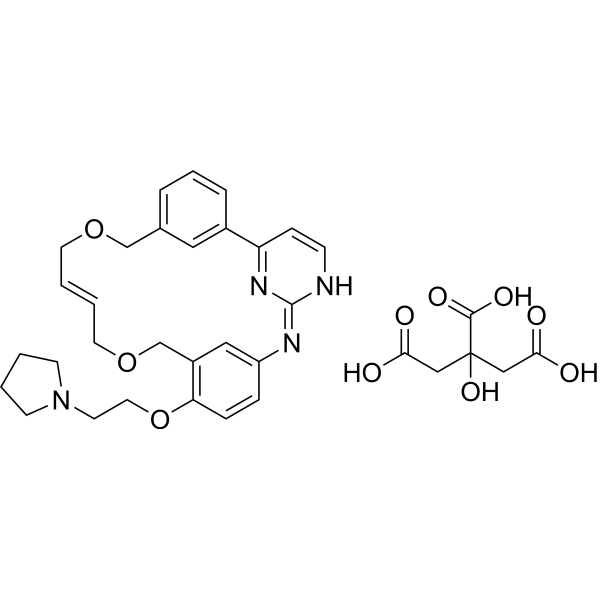
Pacritinib citrate (SB1518 citrate) is an orally active, selective JAK2/FLT3 inhibitor with IC50 values of 23 nM and 22 nM, respectively. Pacritinib citrate inhibits downstream STAT5, ERK1/2, and AKT signaling, and induces Apoptosis and G1 phase arrest. Pacritinib citrate normalizes tumor-induced cytokines/chemokines, and reduces metastasis, tumor growth, shrinkage, migration, liver injury, inflammation, and steatosis. Pacritinib citrate can be used in research on acute myeloid leukemia, myeloproliferative neoplasms, leukemia/lymphoma, GVHD, and alcoholic/non-alcoholic liver fibrosis.
For research use only. We do not sell to patients.
- Purity : 99.68%
- CAS No.: 1228923-42-9
- Formula: C34H40N4O10
- Molecular Weight:664.70
-
Storage:
4°C, sealed storage, away from moisture
* In solvent : -80°C, 6 months; -20°C, 1 month (sealed storage, away from moisture)

To place orders, for customer services and technical support, please contact: MedChemExpress USA
Tel: 609-228-6898 E-mail: [email protected] [email protected]


-
 Biological Activity
-
Chemical Information
-
Solvent & Solubility
- Protocols
- Purity & Documentation
- References
-
 Help & FAQs


Publications Citing Use of MedChemExpress (MCE) Pacritinib citrate
More- Nat Med. 2017 Nov;23(11):1319-1330. [Abstract]
- Science. 2026 Feb 5;391(6785):eadz4075. [Abstract]
- Science. 2017 Dec 1;358(6367):eaan4368. [Abstract]
- Nat Commun. 2021 Aug 13;12(1):4917. [Abstract]
- Nat Commun. 2021 Jul 21;12(1):4441. [Abstract]
- Sci Transl Med. 2018 Jul 18;10(450):eaaq1093. [Abstract]
- Cell Death Dis. 2021 Dec 20;13(1):16. [Abstract]
- Mol Cancer Ther. 2026 Jul 14.
- Mol Syst Biol. 2024 Jan;20(1):28-55. [Abstract]
- Inflamm Res. 2020 Jan;69(1):51-62. [Abstract]
- Drug Des Devel Ther. 2018 Apr 30:12:1009-1017. [Abstract]
- Eur J Pharmacol. 2025 Sep 15:1003:177942. [Abstract]
- PLoS Pathog. 2025 Jun 23;21(6):e1013281. [Abstract]
- PLoS Genet. 2024 May 28;20(5):e1011293. [Abstract]
- BMJ Open Ophthalmol. 2026 Jan 12;11(1):e002307. [Abstract]
- Fundam Clin Pharmacol. 2021 Oct;35(5):919-929. [Abstract]
- Eur J Drug Metab Pharmacokinet. 2021 Sep;46(5):625-635. [Abstract]
- bioRxiv. 2025 June 03.
- bioRxiv. 2025 February 21.
- Patent. US20220323444A1.
 Customer Validation & Images

-
WB
-
RT-PCR
-
In Vivo Efficacy Study
-
Cell Proliferation/Viability Assay
-
WB


Biological Activity
Description
IC50 & Target
[1]|
JAK2 23 nM (IC50) |
FLT3 22 nM (IC50) |
STAT5 |
ERK1 |
ERK2 |
Cellular Effect
|
Cell Line

|
Type | Value | Description | References |
|---|---|---|---|---|
| SET-2 | IC50 |
0.22 μM
|
Antiproliferative activity against human SET-2 cells assessed as viability reduction incubated 48 hrs by CellTiter-Glo assay.
Antiproliferative activity against human SET-2 cells assessed as viability reduction incubated 48 hrs by CellTiter-Glo assay.
|
22829080 |
| MV4-11 | IC50 |
47 nM
|
Antiproliferative activity against human MV4-11 cells assessed as viability reduction incubated 48 hrs by CellTiter-Glo assay.
Antiproliferative activity against human MV4-11 cells assessed as viability reduction incubated 48 hrs by CellTiter-Glo assay.
|
22829080 |
| MOLM-13 | IC50 |
67 nM
|
Antiproliferative activity against human MOLM-13 cells assessed as viability reduction incubated 48 hrs by CellTiter-Glo assay.
Antiproliferative activity against human MOLM-13 cells assessed as viability reduction incubated 48 hrs by CellTiter-Glo assay.
|
22829080 |
| RS4-11 | IC50 |
930 nM
|
Antiproliferative activity against human RS4;11 cells assessed as viability reduction incubated 48 hrs by CellTiter-Glo assay.
Antiproliferative activity against human RS4;11 cells assessed as viability reduction incubated 48 hrs by CellTiter-Glo assay.
|
22829080 |
| MV4-11 | IC50 |
153 nM
|
Antiproliferative activity against human MV4-11-P cells assessed as viability reduction incubated 48 hrs by CellTiter-Glo assay.
Antiproliferative activity against human MV4-11-P cells assessed as viability reduction incubated 48 hrs by CellTiter-Glo assay.
|
22829080 |
| MV4-11 | EC50 |
0.96 μM
|
Induction of apoptosis in human MV4-11 cells assessed as caspase-3/7 activation incubated 16 hrs by Caspase-Glo 3/7 assay.
Induction of apoptosis in human MV4-11 cells assessed as caspase-3/7 activation incubated 16 hrs by Caspase-Glo 3/7 assay.
|
22829080 |
| MV4-11 | IC50 |
80 nM
|
Inhibition of FLT3 autophosphorylation in human MV4-11 cells assessed as pFLT3 (Y591) incubated 3 hrs by immunoblotting.
Inhibition of FLT3 autophosphorylation in human MV4-11 cells assessed as pFLT3 (Y591) incubated 3 hrs by immunoblotting.
|
22829080 |
| MV4-11 | IC50 |
40 nM
|
Inhibition of STAT5 phosphorylation in human MV4-11 cells assessed as pSTAT5 (Y694) incubated 3 hrs by immunoblotting.
Inhibition of STAT5 phosphorylation in human MV4-11 cells assessed as pSTAT5 (Y694) incubated 3 hrs by immunoblotting.
|
22829080 |
| MV4-11 | IC50 |
33 nM
|
Inhibition of ERK1/2 phosphorylation in human MV4-11 cells assessed as pERK1/2 (T202/Y204) incubated 3 hrs by immunoblotting.
Inhibition of ERK1/2 phosphorylation in human MV4-11 cells assessed as pERK1/2 (T202/Y204) incubated 3 hrs by immunoblotting.
|
22829080 |
| MV4-11 | IC50 |
29 nM
|
Inhibition of Akt phosphorylation in human MV4-11 cells assessed as pAkt (T308) incubated 3 hrs by immunoblotting.
Inhibition of Akt phosphorylation in human MV4-11 cells assessed as pAkt (T308) incubated 3 hrs by immunoblotting.
|
22829080 |
| MOLM-13 | IC50 |
180 nM
|
Inhibition of FLT3 autophosphorylation in human MOLM-13 cells assessed as pFLT3 (Y591) after 3 hrs pre-incubation and 3 min FLT3 ligand stimulation by immunoblotting.
Inhibition of FLT3 autophosphorylation in human MOLM-13 cells assessed as pFLT3 (Y591) after 3 hrs pre-incubation and 3 min FLT3 ligand stimulation by immunoblotting.
|
22829080 |
| MOLM-13 | IC50 |
20 nM
|
Inhibition of STAT5 phosphorylation in human MOLM-13 cells assessed as pSTAT5 (Y694) after 3 hrs pre-incubation and 3 min FLT3 ligand stimulation by immunoblotting.
Inhibition of STAT5 phosphorylation in human MOLM-13 cells assessed as pSTAT5 (Y694) after 3 hrs pre-incubation and 3 min FLT3 ligand stimulation by immunoblotting.
|
22829080 |
| RS4-11 | IC50 |
600 nM
|
Inhibition of FLT3 autophosphorylation in human RS4;11 cells assessed as pFLT3 (Y591) after 3 hrs pre-incubation and 3 min FLT3 ligand stimulation by immunoblotting.
Inhibition of FLT3 autophosphorylation in human RS4;11 cells assessed as pFLT3 (Y591) after 3 hrs pre-incubation and 3 min FLT3 ligand stimulation by immunoblotting.
|
22829080 |
| RS4-11 | IC50 |
8 nM
|
Inhibition of STAT5 phosphorylation in human RS4;11 cells assessed as pSTAT5 (Y694) after 3 hrs pre-incubation and 3 min FLT3 ligand stimulation by immunoblotting.
Inhibition of STAT5 phosphorylation in human RS4;11 cells assessed as pSTAT5 (Y694) after 3 hrs pre-incubation and 3 min FLT3 ligand stimulation by immunoblotting.
|
22829080 |
In Vitro
Pacritinib (8-1000 nM; 3 h) citrate inhibits FLT3 autophosphorylation and downstream STAT5, MAPK, and PI3K signaling in FLT3-ITD and FLT3-wt AML cell lines, with pSTAT5 IC50 values of 40 nM in MV4-11, 20 nM in MOLM-13, and 8 nM in RS4;11[1].
Pacritinib citrate inhibits the proliferation of leukemia and lymphoma cell lines dependent on JAK2 or FLT3 activation, with an IC50 of 35-240 nM[4].
Pacritinib citrate inhibits JAK/STAT signaling and proliferation in cancer cell lines, including Karpas 1106P and Karpas1160, with an IC50 of 110 ng/mL for Karpas1160 proliferation[7].
Pacritinib (0.03-0.15 μM; 48-72 h) citrate dose-dependently increases early and late apoptosis in MV4-11 cells without inducing necrosis[1].
Pacritinib citrate combined with Pracinostat (HY-13322) completely eliminates JAK2 autophosphorylation in SET-2 cells and enhances apoptosis markers in SET-2, HEL92.1.7, MV4-11, and MOLM-13 cells[2].
Pacritinib (1 μM; 24 h) citrate reduces fibrosis marker expression and proliferation in primary mouse wild-type and SM22Cre+-Jak2f/f HSCs as well as LX-2 and TWNT-4 HSCs, with JAK2-dependent effects in mouse HSCs[3].
Pacritinib citrate inhibits JAK2, JAK2 V617F, FLT3, and FLT3 D835Y with IC50 values of 6-22 nM[4].
MedChemExpress (MCE) has not independently confirmed the accuracy of these methods. They are for reference only.
-
Cell Line:AML cell lines, including SET-2, MV4-11, MOLM-13, RS4;11, and other AML-derived lines
-
Concentration:up to 10 μM
-
Incubation Time:48 h
-
Result:Showed the highest potency in FAB M5 subtype lines (THP-1, MV4-11, and MOLM-13).
SET-2 (JAK2V617F) IC50 = 0.22 μM; MV4-11 IC50 = 47 nM; MOLM-13 IC50 = 67 nM; RS4;11 IC50 = 930 nM; MV4-11-P IC50 = 153 nM; MV4-11-R IC50 = 434 nM, a 2.9-fold difference between parental and resistant cells.
-
Cell Line:MV4-11 cells
-
Concentration:0.5 nM-10 μM
-
Incubation Time:16 h
-
Result:Dose-dependently activated caspase-3/7, with an EC50 of 0.96 μM.
-
Cell Line:MV4-11 cells
-
Concentration:0.03 and 0.15 μM
-
Incubation Time:48 h and 72 h
-
Result:At 48 h, 0.03 mM increased early and late apoptotic cells to 24.50% and 5.58%, respectively; 0.15 mM increased them to 39.39% and 20.83%.
At 72 h, 0.03 mM increased early and late apoptotic cells to 22.69% and 3.74%; 0.15 mM increased them to 58.69% and 15.10%.
No necrosis induction was observed.
-
Cell Line:MV4-11, MOLM-13, and RS4;11 cells
-
Concentration:8, 40, 200, 1000 nM
-
Incubation Time:3 h (MOLM-13 and RS4;11 plus 10 ng/mL FLT3 ligand for 3 min)
-
Result:In MV4-11, dose-dependent decreases occurred in pFLT3, pSTAT5, pERK1/2, and pAkt with IC50 values of 80, 40, 33, and 29 nM, respectively.
In MOLM-13, IC50 values were 180 nM for pFLT3 and 20 nM for pSTAT5.
In RS4;11, IC50 values were 600 nM for pFLT3 and 8 nM for pSTAT5.
In Vivo
Pacritinib (Pacritinib hydrochloride; 150 mg/kg; oral; twice daily; for 7 consecutive days) citrate inhibits MOLM-13 tumor growth and reduces lung metastasis in the MOLM-13 AML xenograft model in vivo[1].
Pacritinib (oral) citrate demonstrates target inhibition and antitumor activity in the MV4-11 leukemia model in nude mice[4].
MedChemExpress (MCE) has not independently confirmed the accuracy of these methods. They are for reference only.
-
Animal Model:BALB/c nude (BALB/cOlaHsd-Foxn1nu; female; athymic; 9-16 weeks of age at tumor implantation)[1]
-
Dosage:25 mg/kg, 50 mg/kg, and 100 mg/kg (efficacy); 150 mg/kg (target engagement)
-
Administration:oral; once daily; 21 consecutive days; single dose (target engagement)
-
Result:Induced dose-dependent tumor growth inhibition: 38% at 25 mg/kg, 92% at 50 mg/kg, and 121% at 100 mg/kg.
Complete regression was observed in 3/10 mice at 50 mg/kg/day and 8/8 mice at 100 mg/kg/day.
A single 150 mg/kg dose blocked FLT3 and downstream signaling in tumor tissues at 2 and 4 h; pSTAT5 (Y694) was decreased in tumor lysates at 2 and 4 h compared with vehicle.
-
Animal Model:C.B-17/IcrHan (Hsd-Prkdcscid; female; severe combined immunodeficiency)[1]
-
Dosage:150 mg/kg (efficacy); 150 mg/kg (target engagement)
-
Administration:oral; b.i.d.; 7 consecutive days (methods state 8 consecutive days); single dose (target engagement)
-
Result:Dosing resulted in 83% tumor growth inhibition after 7 days of dosing.
Metastatic incidence (mostly in the lungs) was reduced from 0.83 to 0.33 in the high-dose pacritinib group.
A single 150 mg/kg dose blocked FLT3 and downstream signaling at 3 h; in tumor lysates, pSTAT5 (Y694) was fully inhibited 3 h after the last dose on day 7.
-
Animal Model:C.B-17.Cg-PrkdcscidLystbg/Crl (female, 9 weeks old)[2]
-
Dosage:150 mg/kg (doses refer to free base)
-
Administration:p.o. gavage; twice daily; for 19 days (d0-d18)
-
Result:Led to 61% tumor growth inhibition based on tumor volume and 47% tumor growth inhibition based on tumor weight.
pSTAT5 levels in tumors were decreased by about 50% after chronic monotherapy.
No lymph node metastases were observed in mice dosed with pacritinib alone.
Plasma MCP-1 levels in tumor-bearing mice were reduced by 10% after pacritinib monotherapy; IL-6, IP-10, and KC were normalized.
-
Animal Model:C57BL/6J (wild-type, male, 10-12 weeks old)[3]
-
Dosage:65 μg/kg body weight
-
Administration:i.v. injection; twice per week; starting 3 weeks after first CCl4 injection for 4 weeks
-
Result:Reduced serum ALT levels.
Reduced serum AST levels.
Improved liver parenchyma by H&E staining.
Decreased Sirius red collagen staining.
α-SMA immunostaining showed a trend toward reduction.
Collagen-1 and p-moesin protein levels were reduced.
Col1a1 mRNA expression showed a trend toward reduction, while Acta2 and Tgfb showed no changes.
No changes were observed in steatosis markers, oil red O staining, hepatic triglyceride content, or liver-to-body weight ratio.
-
Animal Model:NSG (male or female, age 6 to 24 weeks old)[5]
-
Dosage:50 mg/kg (PAC); 50 mg/kg plus sirolimus (PAC/SIR)
-
Administration:for 14 days
-
Result:Decreased the frequency of CD4+, pSTAT3+ human T cells in mouse spleens at day 14.
Increased the amount of Tregs in mouse spleen at day 14.
Improved survival from xenogeneic GVHD compared with either inhibitor alone.
Reduced GVHD scores.
Decreased the frequency of CD4+, pSTAT3+ human T cells at day 14.
Decreased CD4+, pS6+ T cells at day 14.
Decreased both CD4+, pSTAT3+ and pS6+ T cells.
Decreased the frequency of pathogenic Th1 cells in the spleen at day 14.
Decreased human Th17 cells at day 14.
Had similar Th2 cell frequency.
Did not increase Treg frequency in mice transplanted with standard human PBMCs.
In mice transplanted with Treg-depleted PBMCs, PAC/SIR produced more human Tregs at day 14 in mouse spleens compared with sirolimus.
PAC/SIR-treated mice retained high frequencies of CD4+, pH3ser10+ T cells compared with controls.
- Collagen: Sirius Red Staining
- Annexin V plus membrane-impermeant dye apoptosis staining
- Cell migration
- Connective Tissue: Masson's Trichrome/Collagen Trichrome Staining
- Patient-Derived Orthotopic Xenograft (PDOX)
- Research Protocol for Inflammation-related Diseases
- Liver Histomorphometry
- Fibrosis/Collagen Morphometry
- Detection of Apoptosis
- TUNEL staining for apoptotic DNA fragmentation
- Patient-Derived Xenograft (PDX)
- Apoptosis Solutions
- Subcutaneous Cell-Line-Derived Xenograft
- Orthotopic Cell-Line Xenograft


Chemical Information
-
CAS No. 1228923-42-9
-
Appearance Solid
-
Molecular Weight 664.70
-
Formula C34H40N4O10
-
Color Light yellow to yellow
-
SMILES
OC(CC(CC(O)=O)(C(O)=O)O)=O.C1(COC/C=C/COCC2=C(OCCN3CCCC3)C=CC4=C2)=CC=CC(C5=N/C(NC=C5)=N\4)=C1
-
Synonyms
SB1518 citrate
-
Shipping
Room temperature in continental US; may vary elsewhere.
-
Storage
4°C, sealed storage, away from moisture
* In solvent : -80°C, 6 months; -20°C, 1 month (sealed storage, away from moisture)


Publications (20)
-
Journal Impact Factor
-
Most Recent
-
Nat Med
Chromosome 1q21.3 amplification is a trackable biomarker and actionable target for breast cancer recurrence. [Abstract]2017 Nov;23(11):1319-1330. PMID: 28967919
Pacritinib citrate purchased from MedChemExpress. Usage Cited in: Nat Med. 2017 Nov;23(11):1319-1330. [Abstract]
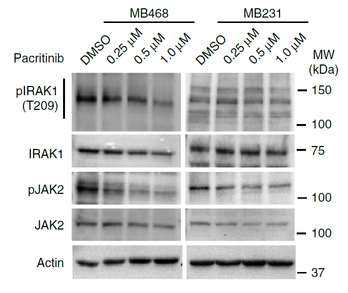
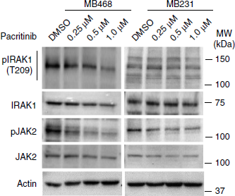
Pacritinib effectively disrupts the S100A7/8/9–IRAK1 feedback loop to inhibit tumorsphere growth. Representative western blot (n=2) showing inhibition of phosphorylated IRAK1 and phosphorylated JAK2 (pJAK2) within 6 h of Pacritinib treatment in MB468 and MB231 cells. Actin is used as a loading control.
-
Science
2026 Feb 5;391(6785):eadz4075. PMID: 41643022 -
Science
2017 Dec 1;358(6367):eaan4368. PMID: 29191878 -
Nat Commun
Genotoxic stress and viral infection induce transient expression of APOBEC3A and pro-inflammatory genes through two distinct pathways. [Abstract]2021 Aug 13;12(1):4917. PMID: 34389714
Pacritinib citrate purchased from MedChemExpress. Usage Cited in: Nat Commun. 2021 Aug 13;12(1):4917. [Abstract]
Pacritinib (0.25-1 µM; 6 h) treatment caused inhibition of phosphorylated IRAK1 and phosphorylated JAK2 (pJAK2) in MB468 and MB231 cells.
Pacritinib citrate purchased from MedChemExpress. Usage Cited in: Nat Commun. 2021 Aug 13;12(1):4917. [Abstract]
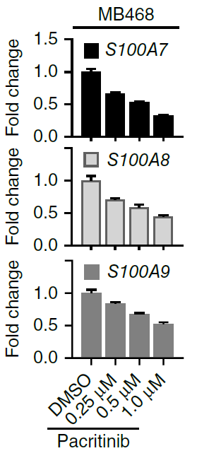
Real-time PCR analysis of S100A7, S100A8, and S100A9 gene expression after Pacritinib (0.25-1 µM; 24 h) treatment in MB468 cells.
Pacritinib citrate purchased from MedChemExpress. Usage Cited in: Nat Commun. 2021 Aug 13;12(1):4917. [Abstract]
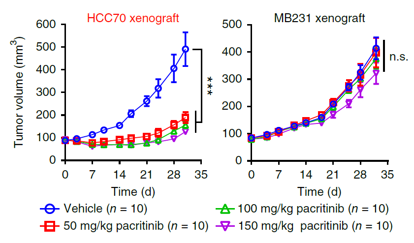
NOD-SCID mice bearing MB231 and HCC70 tumors were treated with pacritinib (50, 100, or 150 mg/kg; p.o.; once daily for 21 d). Pacritinib treatment in different doses resulted in substantial inhibition of tumor growth for HCC70 xenograft tumors but had no obvious effect on the growth of MB231 xenografts.
Pacritinib citrate purchased from MedChemExpress. Usage Cited in: Nat Commun. 2021 Aug 13;12(1):4917. [Abstract]
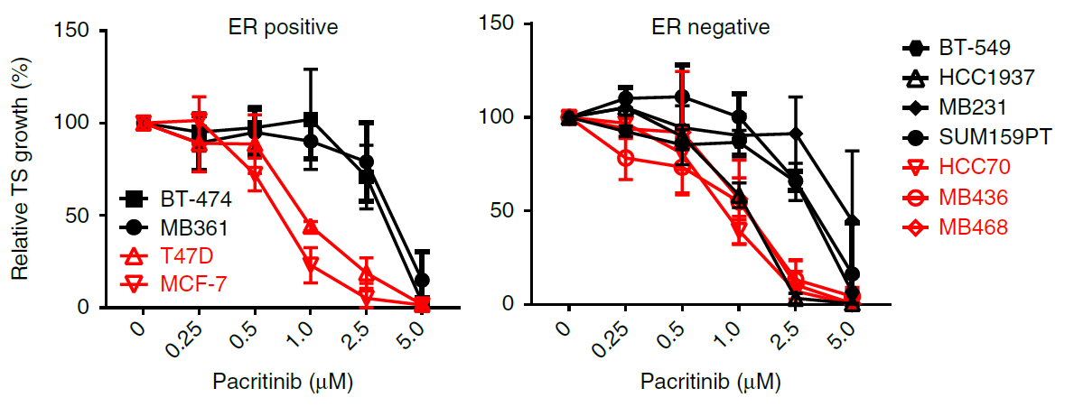
Growth curve of tumorspheres from ER-positive and ER-negative breast cancer cell lines treated with increasing doses of Pacritinib (0.25-5 µM). Pacritinib treatment was more effective in impairment of tumorsphere growth in cancer cell lines positive for 1q21.3 amplification than in cell lines without amplification.
-
Nat Commun
Stromal induction of BRD4 phosphorylation Results in Chromatin Remodeling and BET inhibitor Resistance in Colorectal Cancer. [Abstract]2021 Jul 21;12(1):4441. PMID: 34290255 -
Sci Transl Med
PP2A inhibition is a druggable MEK inhibitor resistance mechanism in KRAS-mutant lung cancer cells. [Abstract]2018 Jul 18;10(450):eaaq1093. PMID: 30021885 -
Cell Death Dis
Colorectal cancer-associated fibroblasts promote metastasis by up-regulating LRG1 through stromal IL-6/STAT3 signaling. [Abstract]2021 Dec 20;13(1):16. PMID: 34930899 -
-
Mol Syst Biol
Illuminating phenotypic drug responses of sarcoma cells to kinase inhibitors by phosphoproteomics. [Abstract]2024 Jan;20(1):28-55. PMID: 38177929 -
Inflamm Res
Potent repression of C-reactive protein (CRP) expression by the JAK1/2 inhibitor ruxolitinib in inflammatory human hepatocytes. [Abstract]2020 Jan;69(1):51-62. PMID: 31654094 -
Drug Des Devel Ther
Advances in the drug therapies of acute myeloid leukemia (except acute wpromyelocytic leukemia). [Abstract]2018 Apr 30:12:1009-1017. PMID: 29750014 -
Eur J Pharmacol
VOPP1, a determinant of the sensitivity of non-small cell lung cancer cells to NAE inhibitors. [Abstract]2025 Sep 15:1003:177942. PMID: 40651787 -
PLoS Pathog
Kaposi sarcoma-associated herpesvirus cooperates with Epstein-Barr virus to co-transform a small set of human B cells oncogenically. [Abstract]2025 Jun 23;21(6):e1013281. PMID: 40549805 -
PLoS Genet
Her2 amplification, Rel-A, and Bach1 can influence APOBEC3A expression in breast cancer cells. [Abstract]2024 May 28;20(5):e1011293. PMID: 38805570 -
BMJ Open Ophthalmol
Impact of anticancer drugs on human Tenon's fibroblast proliferation: implications for glaucoma surgery. [Abstract]2026 Jan 12;11(1):e002307. PMID: 41526033 -
Fundam Clin Pharmacol
2021 Oct;35(5):919-929. PMID: 33523504 -
Eur J Drug Metab Pharmacokinet
Differential Inhibition of Equilibrative Nucleoside Transporter 1 (ENT1) Activity by Tyrosine Kinase Inhibitors. [Abstract]2021 Sep;46(5):625-635. PMID: 34275128 -
-
-


Solvent & Solubility
In Vitro:
DMSO : 30 mg/mL (45.13 mM; ultrasonic and warming and heat to 60°C; Hygroscopic DMSO has a significant impact on the solubility of product, please use newly opened DMSO)
Please refer to the solubility information to select the appropriate solvent. Once prepared, please aliquot and store the solution to prevent product inactivation from repeated freeze-thaw cycles.
Storage method and period of stock solution: -80°C, 6 months; -20°C, 1 month (sealed storage, away from moisture). When stored at -80°C, please use it within 6 months. When stored at -20°C, please use it within 1 month.
Concentration (start) × Volume (start) = Concentration (final) × Volume (final)
In Vivo:
Select the appropriate dissolution method based on your experimental animal and administration route.
- For the following dissolution methods, please ensure to first prepare a clear stock solution using an In Vitro approach and then sequentially add co-solvents:
- To ensure reliable experimental results, the clarified stock solution can be appropriately stored based on storage conditions. As for the working solution for In Vivo experiments, it is recommended to prepare freshly and use it on the same day.
- The percentages shown for the solvents indicate their volumetric ratio in the final prepared solution. If precipitation or phase separation occurs during preparation, heat and/or sonication can be used to aid dissolution.
Add each solvent one by one: 10% DMSO 90% (20% SBE-β-CD in Saline)
Solubility: ≥ 3 mg/mL (4.51 mM); Clear solution
This protocol yields a clear solution of ≥ 3 mg/mL (saturation unknown).
Taking 1 mL working solution as an example, add 100 μL DMSO stock solution (30.0 mg/mL) to 900 μL 20% SBE-β-CD in Saline, and mix evenly.
Preparation of 20% SBE-β-CD in Saline (4°C, storage for one week): 2 g SBE-β-CD powder is dissolved in 10 mL Saline, completely dissolve until clear.
In Vivo Dissolution Calculator
Please enter the basic information of animal experiments:
-
-
-
-
Recommended: Prepare an additional quantity of animals to account for potential losses during experiments.
Please enter your animal formula composition:
-
%DMSO +
Recommended: Keep the proportion of DMSO in working solution below 2% if your animal is weak.
-
%+
-
+%Tween-80 + +
-
%Saline +
The co-solvents required include: DMSO, . All of co-solvents are available by MedChemExpress (MCE). , Tween 80. All of co-solvents are available by MedChemExpress (MCE).
Working solution concentration: 0.22 mg/mL
Method for preparing stock solution: mg drug dissolved in μL DMSO. Stock solution concentration: mg/mL. * In solvent : -80°C, 6 months; -20°C, 1 month (sealed storage, away from moisture)
1. Take μL DMSO stock solution;
2. Add μL .
μL , mix evenly;
3. Then add μL Tween 80, mix evenly;
4. Then add μL
Please ensure that the stock solution in the first step is dissolved to a clear state, and add co-solvents in sequence. You can use ultrasonic heating (ultrasonic cleaner, recommended frequency 20-40 kHz), vortexing, etc. to assist dissolution.


Purity & Documentation
-
Data Sheet (303 KB)
-
SDS (419 KB)
- English - EN (419 KB)
- Français - FR (419 KB)
- Deutsch - DE (419 KB)
- Norwegian - NO (419 KB)
- Español - ES (419 KB)
- Swedish - SV (419 KB)
- Italian - IT (419 KB)
- Korean - KR (419 KB)
- Portuguese - PT (419 KB)
-
Handling Instructions (2659 KB)
References
[1]. Hart S, et al. Pacritinib (SB1518), a JAK2/FLT3 inhibitor for the treatment of acute myeloid leukemia. Blood cancer journal. 2011 Nov;1(11):e44. [Content Brief]
[2]. Novotny-Diermayr V, et al. The oral HDAC inhibitor pracinostat (SB939) is efficacious and synergistic with the JAK2 inhibitor pacritinib (SB1518) in preclinical models of AML. Blood cancer journal. 2012 May;2(5):e69. [Content Brief]


Complete Stock Solution Preparation Table
Please refer to the solubility information to select the appropriate solvent. Once prepared, please aliquot and store the solution to prevent product inactivation from repeated freeze-thaw cycles.
Storage method and period of stock solution: -80°C, 6 months; -20°C, 1 month (sealed storage, away from moisture). When stored at -80°C, please use it within 6 months. When stored at -20°C, please use it within 1 month.
| Optional Solvent | Concentration Solvent Mass | 1 mg | 5 mg | 10 mg | 25 mg |
|---|---|---|---|---|---|
| DMSO | 1 mM | 1.5044 mL | 7.5222 mL | 15.0444 mL | 37.6110 mL |
| 5 mM | 0.3009 mL | 1.5044 mL | 3.0089 mL | 7.5222 mL | |
| 10 mM | 0.1504 mL | 0.7522 mL | 1.5044 mL | 3.7611 mL | |
| 15 mM | 0.1003 mL | 0.5015 mL | 1.0030 mL | 2.5074 mL | |
| 20 mM | 0.0752 mL | 0.3761 mL | 0.7522 mL | 1.8805 mL | |
| 25 mM | 0.0602 mL | 0.3009 mL | 0.6018 mL | 1.5044 mL | |
| 30 mM | 0.0501 mL | 0.2507 mL | 0.5015 mL | 1.2537 mL | |
| 40 mM | 0.0376 mL | 0.1881 mL | 0.3761 mL | 0.9403 mL |


Pacritinib citrate Related Classifications
HY-16379B Related Classifications
 Powered by Bioz