13 Results for "

Electron microscopy

" in MedChemExpress (MCE) Product Catalog:
Products (13)

13 Results for "Electron microscopy" in MCE Product Catalog:

18
18 Publications Verification
Cat. No.: HY-103311
CAS No.: 11103-72-3
Purity:  ≥97.0%
Synonyms: Ammoniated ruthenium oxychloride
Target:  

Calcium Channel

Research Areas:  

Cardiovascular Disease

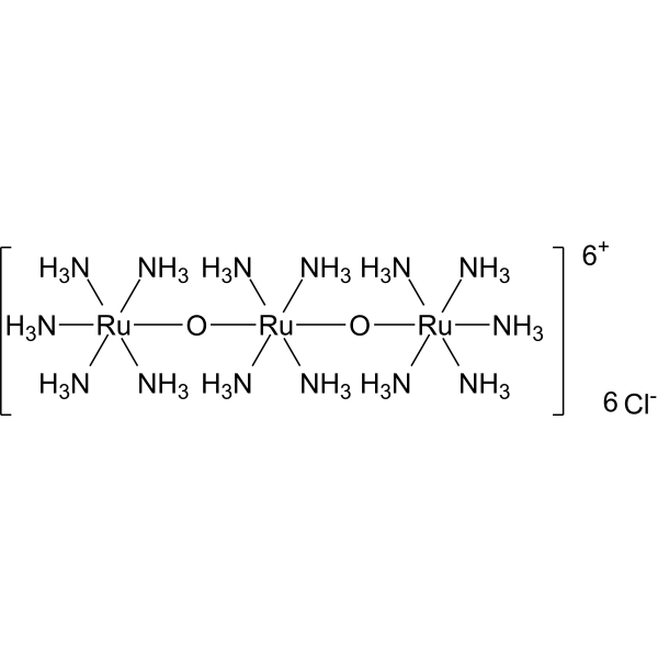
Ruthenium red (Ammoniated ruthenium oxychloride) is a polycationic dye widely used for electron microscopy (EM) of cells, tissues and vegetative bacteria. Ruthenium red strongly reacts with phospholipids and fatty acids and binds to acidic mucopolysaccharides. Ruthenium red is a L-type calcium current (ICa) blocker. When using Ruthenium Red to stain plant tissue sections affected by pathogens, the breakdown of mucilaginous substances by the pathogens prevents them from taking up the stain, whereas the cell walls of healthy tissue stain red .
loading...
    loading...
3
3 Cited Publications
Cat. No.: HY-DY3001
Glutaraldehyde Fixative (2.5%, For Electron Microscopy) is composed of glutaraldehyde, phosphate, deionized water, etc., with a pH range of 7.2-7.4. This fixative is effective in fixing fine structures of cell nuclei and cytoplasm and is commonly used for fixing electron microscopy specimens.
loading...
    loading...
1
1 Cited Publications
Cat. No.: HY-D1668
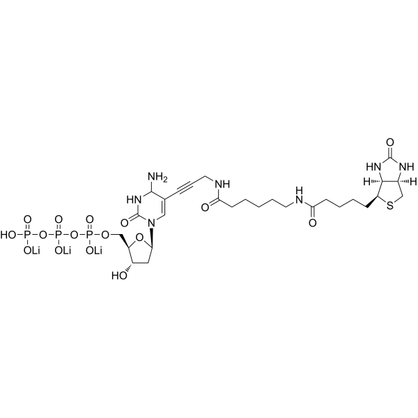
Biotin-11-dCTP is a biotinylated deoxynucleoside triphosphate and an important DNA labeling reagent. In random primer DNA labeling reactions, Biotin-11-dCTP incorporates into newly synthesized DNA strands to generate labeled DNA probes suitable for hybridization applications. In addition, Biotin-11-dCTP can serve as a substrate for terminal deoxynucleotidyl transferase to end-label oligonucleotides for telomere sequence detection, or to label the cut ends of linearized DNA molecules, thereby supporting streptavidin-based electron microscopy analysis. For example, Biotin-11-dCTP can label the cut ends of linearized DNA molecules under the action of dGTP and avian myeloblastosis virus reverse transcriptase .
loading...
    loading...
Cat. No.: HY-DY3002
Glutaraldehyde Fixative (4%, For Electron Microscopy) is composed of glutaraldehyde, phosphate, deionized water, etc., with a pH range of 7.2-7.4. This fixative is effective in fixing fine structures of cell nuclei and cytoplasm and is commonly used for fixing electron microscopy specimens.
loading...
    loading...
Cat. No.: HY-W129441
CAS No.: 91281-32-2
Synonyms: N-Ac-4-S-CAP
Research Areas:  

Others

N-Acetyl-4-S-mercaptoaminophenol (N-Ac-4-S-CAP) is a compound that is selectively cytotoxic to melanocytes of black mouse hair follicles. It can cause 98% depigmentation of black mouse hair follicles. N-Ac-4-S-CAP can produce visible changes in hair follicle melanocytes 4 hours after intraperitoneal injection, including aggregation of melanin granules and nuclear condensation. Electron microscopy observations showed that it caused progressive destruction of melanocytes, including swelling of membranous organelles, nuclear condensation, and cytoplasmic vacuolation, ultimately leading to complete cell necrosis. N-Ac-4-S-CAP has a specific cytotoxic effect on melanocytes that actively produce eumelanin, but may not affect precursor or dormant melanocytes. These properties suggest that N-Ac-4-S-CAP may have potential application value in the treatment of melanoma or skin whitening.
loading...
    loading...
Cat. No.: HY-111137
CAS No.: 916890-10-3
Synonyms: XC-302 free base
Target:  

Akt

Research Areas:  

Cancer

Puquitinib (XC-302 free base) is a multi-target inhibitor with the activity of inducing autophagy in nasopharyngeal carcinoma cells by inhibiting the PI3K/AKT/mTOR signaling pathway. Puquitinib was able to inhibit the proliferation of CNE-2 cells, showing a dose-dependent antiproliferative effect. Puquitinib induced the formation of autophagosomes and autolysosomes in CNE-2 cells, which were observed by fluorescence microscopy and electron microscopy. Puquitinib promoted the formation of LC3-II and increased the expression of beclin 1, while reducing the level of p62. Puquitinib inhibited the PI3K/AKT/mTOR pathway by reducing the expression of p-AKT and p-mTOR. Puquitinib also induced apoptosis in CNE-2 cells, and when autophagy was inhibited, the apoptosis rate was reduced, which means that autophagy may interact with apoptosis to induce cell death .
loading...
    loading...
Cat. No.: HY-173283
Target:  

Fungal

Research Areas:  

Infection

Antifungal agent 129 (Compound 4g) is an inhibitor targeting Rhizoctonia solani, with an EC50 value of 4.27 mg/L. It has a good control effect against rice sheath blight in in vitro and in vivo experiments. Preliminary exploration through scanning electron microscopy, molecular docking, enzyme activity determination, and cytotoxicity experiments has revealed that Antifungal agent 129 may exert its inhibitory activity by affecting the cell structure or physiological processes of Rhizoctonia solani. Antifungal agent 129 can be used in the research of plant diseases in the agricultural field, such as rice sheath blight, which are caused by Rhizoctonia solani .
loading...
    loading...
Cat. No.: HY-W250179
CAS No.: 5168-91-2
Synonyms: Polyoxyethylene (6) cetyl ether
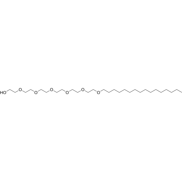
Hexaethylene glycol monohexadecyl ether, is a nonionic surfactant belonging to the polyethylene glycol (PEG) ether family. It has a hydrophilic head and a lipophilic tail, which makes it suitable for a wide range of applications. Specifically, Hexaethylene glycol monohexadecyl ether is commonly used in membrane protein research, for solubilization and stabilization of proteins, and for structural analysis techniques such as X-ray crystallography and electron microscopy. Additionally, Hexaethylene glycol monohexadecyl ether is used in a variety of other industrial and research applications, including drug delivery systems, nanotechnology, and diagnostic analysis. Its unique properties make it ideal for facilitating interactions between molecules with different physicochemical properties.
loading...
    loading...
Cat. No.: HY-DY2047
Research Areas:  

Infection

Phosphotungstic Acid Negative Staining Solution (3%, pH 7.0) is an electron microscopy negative stain. Phosphotungstic Acid Negative Staining Solution (3%, pH 7.0) forms an electron-dense background around samples, enabling the imaging of viruses, bacterial, macromolecules and organelles .
loading...
    loading...
Cat. No.: HY-K3122

MCE Dry Plant-Derived Extracellular Vesicle Isolation and Purification Kit is specifically optimized for dried plant samples. It facilitates the efficient release of vesicles from plant tissues and employs an optimized extraction and purification system to remove non-vesicular components such as polysaccharides and phenolic compounds, thereby enabling the efficient extraction and purification of plant-derived vesicles. The isolated plant-derived vesicles can be used for downstream applications including transmission electron microscopy (TEM), nanoparticle tracking analysis (NTA), Western blotting, qPCR, cell-based studies, and animal experiments.

loading...
    loading...
Cat. No.: HY-K3123

MCE Succulent Plant -Derived Extracellular Vesicle Isolation and Purification Kit is specifically optimized for succulent plant samples. It facilitates the efficient release of vesicles from plant tissues and employs an optimized extraction and purification system to remove non-vesicular components such as polysaccharides and phenolic compounds, thereby enabling the efficient extraction and purification of plant-derived vesicles. The isolated plant-derived vesicles can be used for downstream applications including transmission electron microscopy (TEM), nanoparticle tracking analysis (NTA), Western blotting, qPCR, cell-based studies, and animal experiments.

loading...
    loading...
Cat. No.: HY-K3124

MCE Macrofungal-Derived Extracellular Vesicle Isolation and Purification Kit is specifically optimized for fungal fruiting body samples. The optimized extraction system facilitates the release of EVs from fungal tissues, while the purification system effectively removes polysaccharides, proteins, phenolic compounds, terpenoids, and other non-vesicular components, enabling efficient isolation and purification of fungal EVs. The isolated macrofungal-derived vesicles can be used for downstream applications including transmission electron microscopy (TEM), nanoparticle tracking analysis (NTA), Western blotting, qPCR, cell-based studies, and animal experiments.

loading...
    loading...
Cat. No.: HY-DY2034
Target:  

Calcium Channel

Research Areas:  

Cardiovascular Disease

Ruthenium Red Staining Solution (0.01%) is a polycationic dye widely used for electron microscopy (EM) of cells, tissues and vegetative bacteria. Ruthenium Red Staining Solution (0.01%) strongly reacts with phospholipids and fatty acids and binds to acidic mucopolysaccharides. Ruthenium Red Staining Solution (0.01%) is a L-type calcium current (ICa) blocker. When using Ruthenium Red Staining Solution (0.01%) to stain plant tissue sections affected by pathogens, the breakdown of mucilaginous substances by the pathogens prevents them from taking up the stain, whereas the cell walls of healthy tissue stain red .
loading...
    loading...
  • 1