263 Results for "

microscopy

" in MedChemExpress (MCE) Product Catalog:
Products (263)

263 Results for "microscopy" in MCE Product Catalog:

191
191 Publications Verification
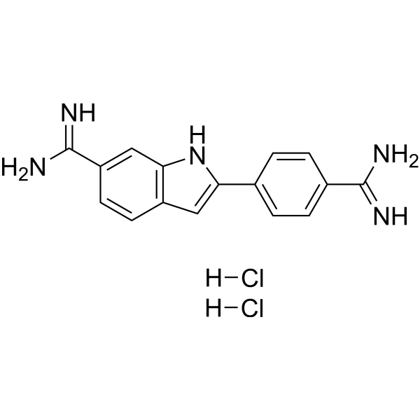
Cat. No.: HY-D0814
CAS No.: 28718-90-3
Synonyms: 4',6-Diamidino-2-phenylindole dihydrochloride
DAPI (4',6-Diamidino-2-phenylindole) dihydrochloride is a DAPI dye. DAPI is a fluorescent dye that binds strongly to DNA. It binds to the AT base pair of the double-stranded DNA minor groove, and one DAPI molecule can occupy three base pair positions. The fluorescence intensity of DAPI molecules bound to double-stranded DNA is increased by about 20 times, and it is commonly observed with fluorescence microscopy, and the amount of DNA can be determined based on the intensity of fluorescence. DAPI cannot penetrate intact cell membranes and is commonly used for staining damaged and fixed cells . DAPI (Compound 3) is an acid-sensing ion channel 3 (ASIC3) inhibitor. DAPI binds to ASIC3 and blocks the channel function. DAPI can be used in the study of chronic pain treatment (Ex/Em = 356/451 nm).
loading...
    loading...
191
191 Publications Verification
Cat. No.: HY-D2868
CAS No.: 47165-04-8
Synonyms: 4',6-Diamidino-2-phenylindole
DAPI (4',6-Diamidino-2-phenylindole) is a DAPI dye. DAPI is a fluorescent dye that binds strongly to DNA. It binds to the AT base pair of the double-stranded DNA minor groove, and one DAPI molecule can occupy three base pair positions. The fluorescence intensity of DAPI molecules bound to double-stranded DNA is increased by about 20 times, and it is commonly observed with fluorescence microscopy, and the amount of DNA can be determined based on the intensity of fluorescence. DAPI cannot penetrate intact cell membranes and is commonly used for staining damaged and fixed cells . DAPI (Compound 3) is an acid-sensing ion channel 3 (ASIC3) inhibitor. DAPI binds to ASIC3 and blocks the channel function. DAPI can be used in the study of chronic pain treatment (Ex/Em = 356/451 nm).
loading...
    loading...
185
185 Cited Publications
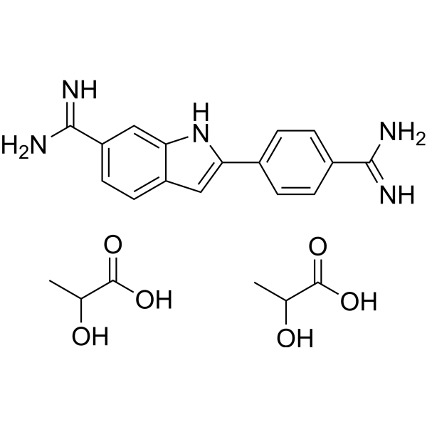
Cat. No.: HY-D1738
CAS No.: 28718-91-4
Synonyms: 4',6-Diamidino-2-phenylindole dilactate
DAPI (4',6-Diamidino-2-phenylindole) dilactate is a DAPI dye. DAPI is a fluorescent dye that binds strongly to DNA. It binds to the AT base pair of the double-stranded DNA minor groove, and one DAPI molecule can occupy three base pair positions. The fluorescence intensity of DAPI molecules bound to double-stranded DNA is increased by about 20 times, and it is commonly observed with fluorescence microscopy, and the amount of DNA can be determined based on the intensity of fluorescence. DAPI cannot penetrate intact cell membranes and is commonly used for staining damaged and fixed cells . DAPI (Compound 3) is an acid-sensing ion channel 3 (ASIC3) inhibitor. DAPI binds to ASIC3 and blocks the channel function. DAPI can be used in the study of chronic pain treatment (Ex/Em = 356/451 nm) .
loading...
    loading...
139
139 Cited Publications
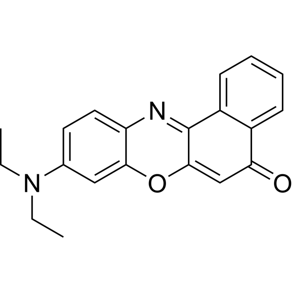
Cat. No.: HY-D0718
CAS No.: 7385-67-3
Synonyms: Nile Blue A oxazone; Phenoxazone 9
Target:  

Fluorescent Dye

Research Areas:  

Others

Nile red (Nile blue oxazone) is a lipophilic stain. Nile red has environment-sensitive fluorescence. Nile red is intensely fluorescent in a lipid-rich environment while it has minimal fluorescence in aqueous media. Nile red is an excellent vital stain for the detection of intracellular lipid droplets by fluorescence microscopy and flow cytof uorometry. Nile red stains intracellular lipid droplets red. The fluorescence wavelength is 543/598 nm .
loading...
    loading...
86
86 Cited Publications
Cat. No.: HY-116215
CAS No.: 186689-07-6
Purity:  99.74%
Target:  

Fluorescent Dye

Research Areas:  

Others

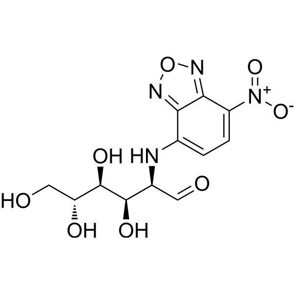
2-NBDG is a fluorescently-labeled deoxyglucose analog that is used primarily to directly monitor glucose uptake by living cells and tissues. It is also used as a topical contrast reagent for the detection of neoplasia. 2-NBDG can be used in real-time confocal, high-resolution, or wide-field fluorescence microscopy as well as in flow cytometry. The probe can be excited by the Argon laser at 488 nm to give the environment-sensitive fluorescence. It has lower photostability than the rhodamine-based fluorescent probes.
loading...
    loading...
44
44 Cited Publications
Cat. No.: HY-D0952
CAS No.: 494-38-2
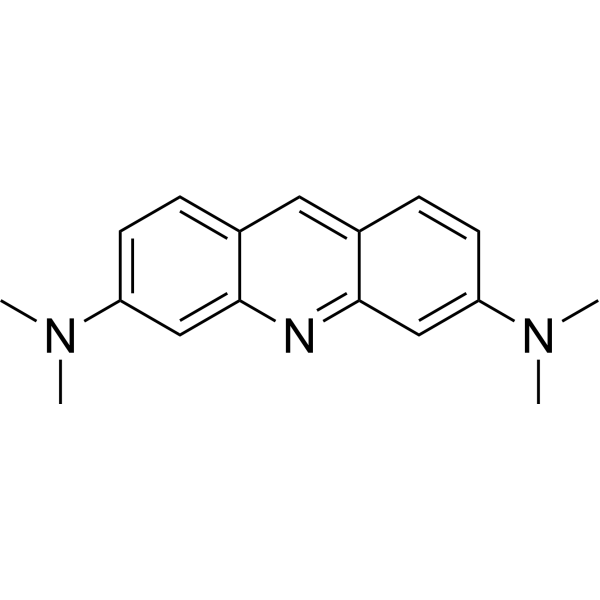
Acridine Orange base is a cell-permeable fluorescent dye that stains organisms (bacteria, parasites, viruses, etc.) bright orange and, when used under appropriate conditions (pH=3.5, Ex=460 nm), distinguishes human cells in green for detection by fluorescence microscopy. Acridine Orange base fluoresces green when bound to dsDNA (Ex=488, Em=520-524) and red when bound to ssDNA (Ex=457, Em=630-644) or ssRNA (Ex=457, Em=630-644), also can be used in cell cycle assays .
loading...
    loading...
21
21 Cited Publications
Cat. No.: HY-D0090
CAS No.: 162558-52-3
Target:  

Fluorescent Dye

Research Areas:  

Others

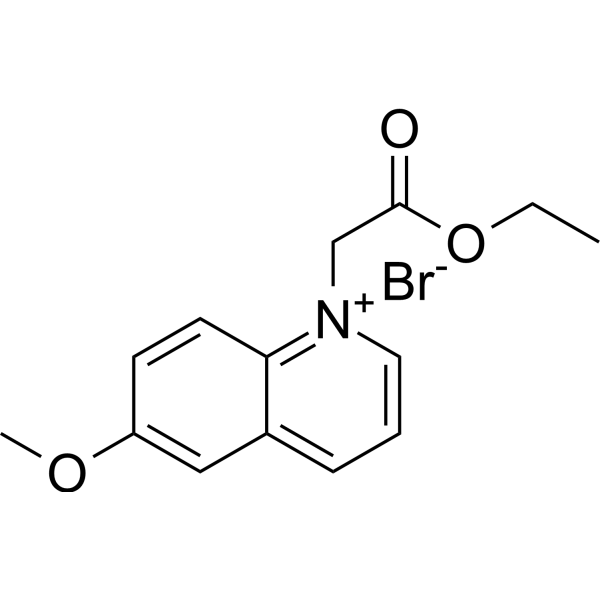
MQAE is a chloride ion (Cl -) fluorescent probe that can be used to measure chloride concentrations. The fluorescence intensity of MQAE decreases proportionally as Cl - ions increase. MQAE has high cell permeability and is suitable for fluorescence detection such as confocal microscopy and flow cytometry (Ex/Em=350/460 nm) .
loading...
    loading...
18
18 Cited Publications
Cat. No.: HY-103311
CAS No.: 11103-72-3
Purity:  ≥97.0%
Synonyms: Ammoniated ruthenium oxychloride
Target:  

Calcium Channel

Research Areas:  

Cardiovascular Disease

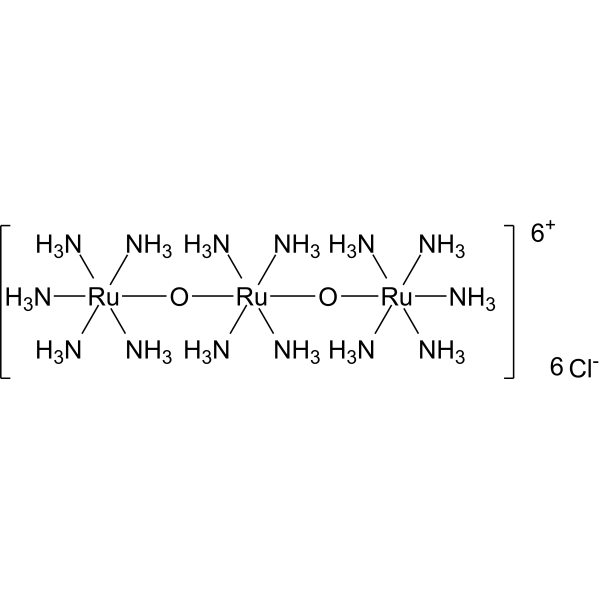
Ruthenium red (Ammoniated ruthenium oxychloride) is a polycationic dye widely used for electron microscopy (EM) of cells, tissues and vegetative bacteria. Ruthenium red strongly reacts with phospholipids and fatty acids and binds to acidic mucopolysaccharides. Ruthenium red is a L-type calcium current (ICa) blocker. When using Ruthenium Red to stain plant tissue sections affected by pathogens, the breakdown of mucilaginous substances by the pathogens prevents them from taking up the stain, whereas the cell walls of healthy tissue stain red .
loading...
    loading...
14
14 Cited Publications
Cat. No.: HY-111330
CAS No.: 359010-69-8
Purity:  99.91%
Synonyms: HPF; 3'-p-(Hydroxyphenyl) fluorescein
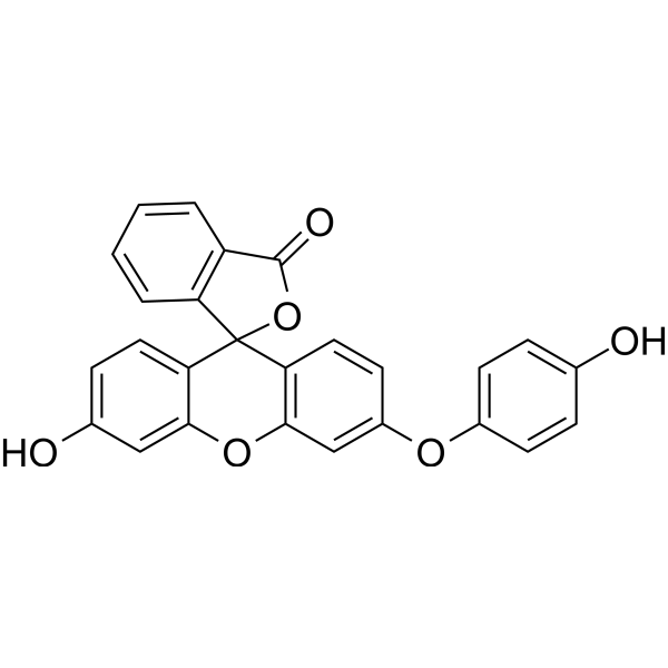
Hydroxyphenyl Fluorescein (HPF) is a stable ROS fluorescent probe dye. Hydroxyphenyl Fluorescein has stronger specificity and stability than H2DCFDA (HY-D0940). Hydroxyphenyl Fluorescein can produce strong green fluorescence through hydroxyl radical reaction with intracellular peroxynitroso. Hydroxyphenyl Fluorescein can be applied for fluorescence microscopy, high-throughput imager, luciferase microplate reader or flow cytometry. Ex/Em=490/515 nm .
loading...
    loading...
3
3 Cited Publications
Cat. No.: HY-DY3001
Glutaraldehyde Fixative (2.5%, For Electron Microscopy) is composed of glutaraldehyde, phosphate, deionized water, etc., with a pH range of 7.2-7.4. This fixative is effective in fixing fine structures of cell nuclei and cytoplasm and is commonly used for fixing electron microscopy specimens.
loading...
    loading...
3
3 Cited Publications
Cat. No.: HY-D1396
CAS No.: 2387906-44-5
Research Areas:  

Others

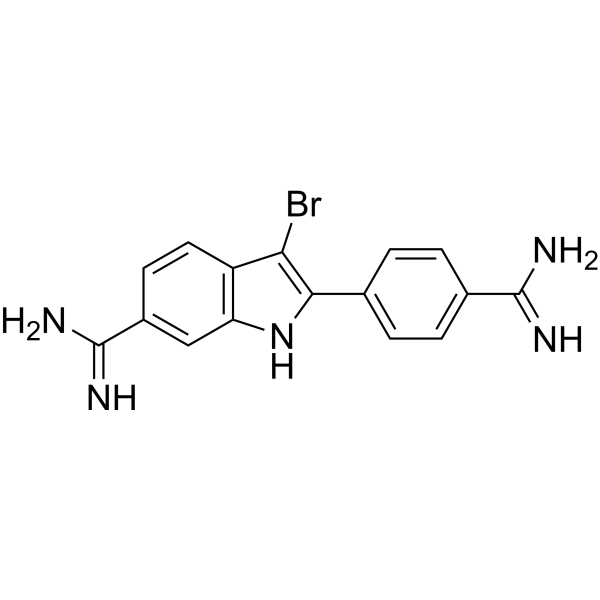
Br-DAPI is a marker dye in DAPI series. DAPI is a fluorescent dye that binds strongly to DNA. It binds to the AT base pair of the double-stranded DNA minor groove, and one DAPI molecule can occupy three base pair positions. The fluorescence intensity of DAPI molecules bound to double-stranded DNA is increased by about 20 times, and it is commonly observed with fluorescence microscopy, and the amount of DNA can be determined based on the intensity of fluorescence. DAPI cannot penetrate intact cell membranes and is commonly used for staining damaged and fixed cells . Storage: Keep away from light.
loading...
    loading...
1
1 Cited Publications
Cat. No.: HY-D0988
CAS No.: 11016-17-4
Synonyms: R-PE
R-Phycoerythrin is found in Heterosiphonia japonica. R-Phycoerythrin is an orange-red fluorescent probe with α, β, and γ subunits. R-Phycoerythrin can be used in photodynamic therapy (PDT) to induce apoptosis in tumor cells. R-Phycoerythrin can be used in fluorescence microscopy, flow cytometry, and immunofluorescence analysis (Ex: 495 nm).
loading...
    loading...
1
1 Cited Publications
Cat. No.: HY-146245F
Purity:  98.03%
Research Areas:  

Inflammation/Immunology Cancer

FAM-labeled ODN 1826 sodium, a class B CpG ODN (oligodeoxynucleotide), is a TLR9 agonist. FAM-labeled ODN 1826 sodium can be used to evaluate CpG ODN cellular uptake and localization by confocal laser-scanning microscopy or flow cytometry.
loading...
    loading...
1
1 Cited Publications
Cat. No.: HY-W250150
CAS No.: 5486-84-0
Research Areas:  

Others

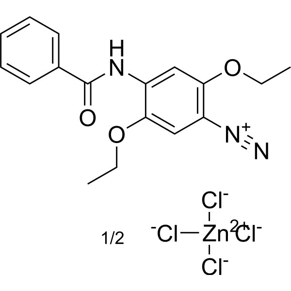
Fast blue bb salt (hemi(zinc chloride) salt is a water-soluble dye commonly used as a histological stain in various biological applications such as staining cellular components or detecting enzyme activity. Fast blue BB salt has a unique Chemical properties that allow it to bind to specific proteins to form a blue product that can be detected by microscopy or spectrophotometry. This makes it a useful tool for visualizing and quantifying target proteins in tissue samples or cell cultures.
loading...
    loading...
1
1 Cited Publications
Cat. No.: HY-D1540
CAS No.: 2097714-45-7
Synonyms: Cy 5.5 amine; Lumiprobe Cy 5.5 amine
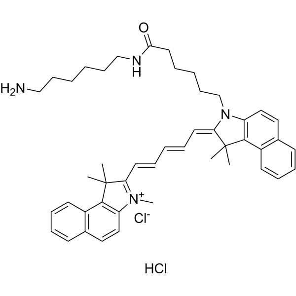
Cyanine5.5 amine (Cy 5.5 amine), a Cy5.5 Analogue, is a near-infrared (NIR) fluorescent dye (Ex=648 nm, Em=710 nm). Cyanine5.5 amine can be used in the preparation of Cy5.5-labeled nanoparticles, which can be tracked and imaged with low fluorescence background using confocal microscopy .
loading...
    loading...
1
1 Cited Publications
Cat. No.: HY-P2031
CAS No.: 26645-35-2
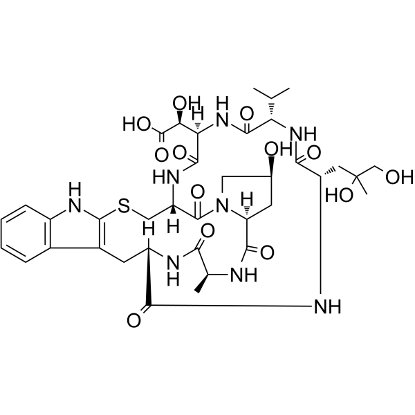
Phallacidin is a natural bicyclic heptapeptide derived from the poisonous mushroom Amanita phalloides. Phallacidin binds to filamentous actin specifically with high affinity, with a Kd of 20 nM. After binding to F-actin, Phallacidin strongly inhibits its depolymerization, stabilizes microfilament structures, and prevents their disruption by drugs such as cytochalasins. When conjugated with a fluorophore, Phallacidin serves as a specific fluorescent probe for F-actin, which is used to clearly visualize the distribution of actin in the cytoskeleton (e.g., stress fibers, cortical peripheral bands) under fluorescence microscopy .
loading...
    loading...
1
1 Cited Publications
Cat. No.: HY-D0163
CAS No.: 14855-76-6
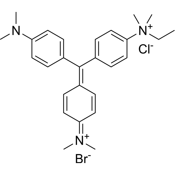
Methyl Green is a non-intercalating fluorescent labeling agent that selectively binds to the major groove of DNA. Methyl Green electrostatically interacts with the major groove of DNA through positively charged groups, exhibiting key activities such as high affinity, resistance to photobleaching, and stable fluorescence emission. Methyl Green can be directly measured by microscopy and flow cytometry, with peaks at 633 and 677 nm. Methyl Green can be used for fluorescent labeling of the nuclei of embryonic tissues or cells, or DNA staining and cell activity detection in gel electrophoresis[1][2][3].
loading...
    loading...
1
1 Cited Publications
Cat. No.: HY-133535
CAS No.: 2093120-32-0
Synonyms: PA-JF646-NHS
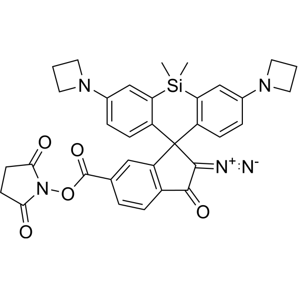
PA Janelia Fluor 646, SE (PA-JF646-NHS) is a photo-activatable fluorescent dye. PA Janelia Fluor 646, SE is a far-red light-excited, 405 nm-activated NHS ester-based dye. Janelia Fluor fluorescent dyes can be chemically conjugated to the specific HaloTag substrate (a chloroalkane ligand) to form a fluorescent ligand, rather than binding directly to the HaloTag protein itself. PA Janelia Fluor 646, SE can achieve extremely low background and single-molecule-level ultra-high-resolution imaging through photo-controlled switching, making it a powerful tool for live cell super-resolution microscopy technology (Ex/Em = 646/664 nm).
loading...
    loading...
1
1 Cited Publications
Cat. No.: HY-NP163A
Synonyms: WGA-AF488
Wheat germ agglutinin-AF488 (WGA-AF488) is a cell membrane-specific staining agent prepared by conjugating wheat germ agglutinin with the Alexa Fluor 488 (HY-D1304) fluorescent dye, and it binds to cell surface glycoproteins with high affinity. Wheat germ agglutinin-AF488 is applied in fluorescence microscopy and confocal imaging techniques, and it can clearly label the membrane structures of various cells including breast cancer cells, enabling high-resolution visual observation. Wheat germ agglutinin-AF488 is used in studies of breast cancer and triple-negative breast cancer to observe cell morphology and membrane dynamic changes .
loading...
    loading...
1
1 Cited Publications
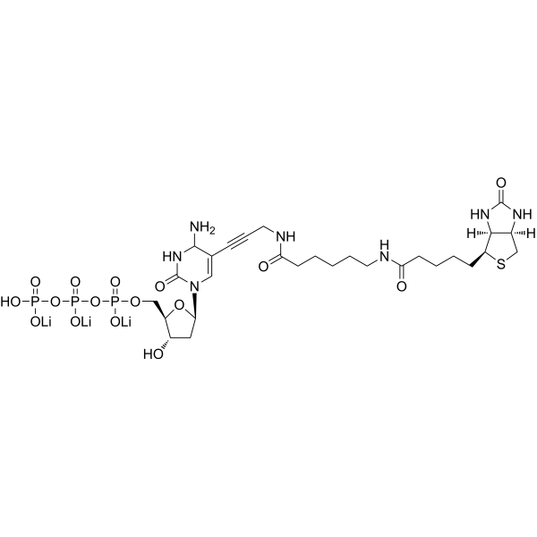
Cat. No.: HY-D1668
Biotin-11-dCTP is a biotinylated deoxynucleoside triphosphate and an important DNA labeling reagent. In random primer DNA labeling reactions, Biotin-11-dCTP incorporates into newly synthesized DNA strands to generate labeled DNA probes suitable for hybridization applications. In addition, Biotin-11-dCTP can serve as a substrate for terminal deoxynucleotidyl transferase to end-label oligonucleotides for telomere sequence detection, or to label the cut ends of linearized DNA molecules, thereby supporting streptavidin-based electron microscopy analysis. For example, Biotin-11-dCTP can label the cut ends of linearized DNA molecules under the action of dGTP and avian myeloblastosis virus reverse transcriptase .
loading...
    loading...