3 Results for "

time-lapse image

" in MedChemExpress (MCE) Product Catalog:
Products (3)

3 Results for "time-lapse image" in MCE Product Catalog:

Cat. No.: HY-D2465
Target:  

Fluorescent Dye

Research Areas:  

Infection

CY5-Dextran (MW60-80k) is a fluorescent probe formed by covalent conjugation of the cyanine dye Cy5 to dextran. It is a pH-insensitive fluid-phase endocytosis tracer (the pH of endosomal lumen is precisely quantified by the fluorescence ratio of FITC/Cy5). CY5-Dextran (MW60-80k) is internalized by cells via a clathrin-independent fluid-phase pinocytosis pathway, and is sequentially evidenced by early endosomes, late endosomes are transported to lysosomes. CY5-D I cannot pass through the limited pore size formed by uncoating of human rhinovirus type 2 (HRV2), but can be released into the neutral cytoplasm upon adenovirus-mediated endosomal rupture, thus distinguishing between the two viral modes of "pore-forming escape" and "lysis escape". CY5-Dextran (MW60) -80k) has an excitation wavelength of 635 nm, and it can also be used for time-lapse imaging of endocytosis dynamics of stromal cells in tissues such as rat salivary glands under in vivo two-photon microscopy .
loading...
    loading...
Cat. No.: HY-D1460
CAS No.: 134869-04-8
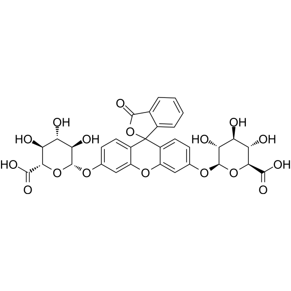
Fluorescein Di-β-D-Glucuronide is an orally active fluorescent probe substrate and an indicator of β-glucuronidase (βG) activity in intestinal bacteria. Fluorescein Di-β-D-Glucuronide can be used for non-invasive time-lapse optical imaging of βG activity in intestinal bacteria in nude mice. Fluorescein Di-β-D-Glucuronide supports the quantitative analysis of bacterial βG enzyme activity and inhibition .
loading...
    loading...
Cat. No.: HY-D3197
CDg16 is a selective fluorescent dye targeting SLC18B1 (λabs/λem=458/544 nm) that is actively transported into lysosomal vesicles of activated macrophages independent of the endocytic pathway. CDg16 enables highly specific vesicle localization in live cells. CDg16 exhibits no cytotoxicity and accurately distinguishes activated M1 and M2 subsets from different origins. CDg16 shows low background staining in non-activated cells and normal organs, making it suitable for time-lapse imaging. In preclinical animal models of inflammatory sites, atherosclerotic plaques and liver inflammation, CDg16 allows visualization of activated macrophages. CDg16 can be used to study inflammation-related diseases and atherosclerosis .
loading...
    loading...