



SGI-1776
Based on 12 publication(s) in Google Scholar
SGI-1776 is an orally active Pim kinase and FLT3 inhibitor with IC50 values of 7 nM, 363 nM, 69 nM, and 44 nM for Pim-1, Pim-2, Pim-3, and FLT3, respectively. SGI-1776 downregulates C/EBP-α, PPAR-γ, FAS, Leptin, and RANTES, and decreases STAT-3 phosphorylation. SGI-1776 induces Apoptosis, cell cycle arrest, mitochondrial depolarization, and reduces RNA synthesis, protein synthesis, and Mcl-1. SGI-1776 inhibits adipogenesis and lipid accumulation. SGI-1776 has anticancer activity against leukemia.
For research use only. We do not sell to patients.
- CAS No.: 1173928-26-1
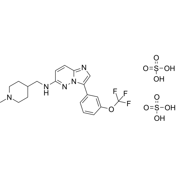
- Formula: C20H26F3N5O9S2
- Molecular Weight:601.57
-
Storage:
Please store the product under the recommended conditions in the Certificate of Analysis.

To place orders, for customer services and technical support, please contact: MedChemExpress USA
Tel: 609-228-6898 E-mail: [email protected] [email protected]


-
 Biological Activity
-
Chemical Information
- Protocols
- Purity & Documentation
- References
-
 Help & FAQs


Publications Citing Use of MedChemExpress (MCE) SGI-1776
More- Science. 2017 Dec 1;358(6367):eaan4368. [Abstract]
- Sci Transl Med. 2018 Jul 18;10(450):eaaq1093. [Abstract]
- Adv Sci (Weinh). 2025 Jul 6:e03487. [Abstract]
- Cell Death Dis. 2018 Feb 22;9(3):307. [Abstract]
- Phytomedicine. 2026 May 15:157:158301. [Abstract]
- Cell Chem Biol. 2024 Feb 15;31(2):326-337.e11. [Abstract]
- J Med Chem. 2021 Nov 11;64(21):15702-15715. [Abstract]
- Front Cell Infect Microbiol. 2024 Nov 29:14:1502770. [Abstract]
- Food Chem Toxicol. 2026 Nov:217:116309.
- eNeuro. 2019 Aug 22;6(4):ENEURO.0003-19.2019. [Abstract]
- Anticancer Drugs. 2024 Jan 1;35(1):46-54. [Abstract]
- Patent. US20180263995A1.
 Customer Validation & Images

-
WB
All DNA/RNA Synthesis Isoforms
More

Biological Activity
Description
|
Mcl-1 |
PIM1 7 nM (IC50) |
PIM2 363 nM (IC50) |
PIM3 69 nM (IC50) |
PPAR-γ |
Stat-3 |
Cellular Effect
|
Cell Line

|
Type | Value | Description | References |
|---|---|---|---|---|
| MOLM-13 | IC50 |
10 nM
|
Viability IC50 of less than 10 nM against human FLT3-ITD AML cell line MOLM-13 after 72 hrs incubation with SGI-1776, associated with apoptosis induction.
Viability IC50 of less than 10 nM against human FLT3-ITD AML cell line MOLM-13 after 72 hrs incubation with SGI-1776, associated with apoptosis induction.
|
561980 |
| MV4-11 | IC50 |
10 nM
|
Viability IC50 of less than 10 nM against human FLT3-ITD AML cell line MV4-11 after 72 hrs incubation with SGI-1776, associated with apoptosis induction.
Viability IC50 of less than 10 nM against human FLT3-ITD AML cell line MV4-11 after 72 hrs incubation with SGI-1776, associated with apoptosis induction.
|
561980 |
| JeKo-1 | IC50 |
5 μM
|
Inhibition of RNA synthesis in human mantle cell lymphoma Jeko cells assessed by uridine incorporation with 50% reduction after exposure to SGI-1776.
Inhibition of RNA synthesis in human mantle cell lymphoma Jeko cells assessed by uridine incorporation with 50% reduction after exposure to SGI-1776.
|
24290221 |
| Mino | IC50 |
10 μM
|
Inhibition of RNA synthesis in human mantle cell lymphoma Mino cells assessed by uridine incorporation with 50% reduction after exposure to SGI-1776.
Inhibition of RNA synthesis in human mantle cell lymphoma Mino cells assessed by uridine incorporation with 50% reduction after exposure to SGI-1776.
|
24290221 |
In Vitro
SGI-1776 (3-10 µM; 24 h) inhibits clonogenic survival of MV-4-11 cells, reducing colony formation to approximately 5% of control at 10 µM[2].
SGI-1776 (2.5-5 μM; 72 h) reduces colony formation in SACC-83 and SACC-LM cells[3].
SGI-1776 inhibits the proliferation of mouse ALL and hALL cell lines, with IC50 values of approximately 1 mM and 2.5 mM, respectively[5].
SGI-1776 (0.1-10 µM; 24 hours) selectively reduces Mcl-1 protein levels in MV-4-11, MOLM-13, and OCI-AML-3 AML cell lines without altering Bcl-2, Bcl-xL, Bak, or Bax[2].
SGI-1776 reduces the viability and clonogenic survival of 9 hAML cell lines, inhibits proliferation and induces apoptosis, while decreasing Bad phosphorylation and increasing p27 expression[7].
SGI-1776 (1-10 μM; 24 h) induces apoptosis in primary AML cells, with an average increase of 5-19%[8].
SGI-1776 (2.5-5 μM; 72 h) induces early apoptosis in SACC-83 and SACC-LM cells in a dose-dependent manner[3].
SGI-1776 (2.5-5 μM; 72 h) increases caspase-3 activity in SACC-83 and SACC-LM cells, with higher activity in SACC-LM cells[3].
SGI-1776 (5-10 µM; 8 days of differentiation) reduces triglyceride content in differentiating 3T3-L1 adipocytes, with the greatest reduction at 10 µM and activity at 5 µM[1].
SGI-1776 (5-10 µM; 8-day differentiation) does not significantly reduce 3T3-L1 adipocyte viability at 5 µM after 8 days, whereas 10 µM causes a slight decrease[1].
SGI-1776 (5 µM; days 0-8) inhibits 3T3-L1 adipogenesis mainly when present during the early differentiation stage, and incubation on days 0-8 produces the highest inhibition rate[1].
MedChemExpress (MCE) has not independently confirmed the accuracy of these methods. They are for reference only.
-
Cell Line:3T3-L1 preadipocytes differentiated into mature adipocytes
-
Concentration:5 and 10 µM
-
Incubation Time:8-day differentiation
-
Result:SGI-1776 for 8 days markedly reduced lipid droplets in a concentration-dependent manner compared with mock control cells.
10 µM SGI-1776 completely blocked accumulation of lipid droplets.
Light microscopy confirmed the SGI-1776-mediated lipid-reducing effect.
SGI-1776 at 5 and 10 µM reduced Oil Red O staining relative to control.
-
Cell Line:3T3-L1 adipocytes
-
Concentration:5 and 10 µM
-
Incubation Time:8-day differentiation
-
Result:Induced a slight decrease in viability at 10 µM, but not at 5 µM.
SGI-1776 at 5 µM for 8 days did not markedly affect the number of 3T3-L1 adipocytes.
-
Cell Line:3T3-L1 preadipocytes/adipocytes
-
Concentration:5 µM
-
Incubation Time:days 2, 5, 8 during differentiation; 2, 4, 8, 24 h after MDI induction
-
Result:Markedly inhibited adipogenesis-dependent protein expression of C/EBP-α and PPAR-γ during adipocyte differentiation, particularly on days 5 and 8.
Markedly decreased phosphorylation levels of STAT-3 on day 2 during adipocyte differentiation.
Did not markedly affect phosphorylation levels of STAT-5.
Suppressed STAT-3 phosphorylation during early stages of adipogenesis induced by MDI at 2, 4, 8 and 24 h.
Did not markedly alter total protein levels of STAT-3 and STAT-5 during adipocyte differentiation.
Markedly suppressed FAS protein expression on days 5 and 8.
-
Cell Line:MV-4-11, MOLM-13, and OCI-AML-3
-
Concentration:0.1, 0.3, 1, 3, 10 µM; DMSO vehicle control
-
Incubation Time:24 h
-
Result:Induced a dose-dependent increase in annexin/PI-positive cells in MV-4-11, MOLM-13, and OCI-AML-3 cells.
In MV-4-11, annexin/PI-positive cells were 6% at 0 µM, 6% with DMSO, 13% at 0.1 µM, 20% at 0.3 µM, 26% at 1 µM, 28% at 3 µM, and 75% at 10 µM.
MOLM-13 and OCI-AML-3 showed dose-dependent increases in annexin/PI-positive cells.
Higher apoptosis occurred in MV-4-11 and MOLM-13, which are FLT3-ITD positive.
In Vivo
SGI-1776 (200 mg/kg/dose; oral; daily × 5 per week) shows in vivo activity in ALL xenografts, reducing tumor proliferation compared with untreated mice[5].
MedChemExpress (MCE) has not independently confirmed the accuracy of these methods. They are for reference only.
-
Animal Model:NOD-SCID (female)[2]
-
Dosage:50 mg/kg (daily 5 days/week for 3 weeks); 75 mg/kg (daily 5 days/week for 3 weeks); 100 mg/kg (twice weekly for 3 weeks); 200 mg/kg (twice weekly for 3 weeks)
-
Administration:p.o.; daily 5 days/week for 3 weeks (50, 75 mg/kg); twice weekly for 3 weeks (100, 200 mg/kg)
-
Result:In MV-4-11 xenografts, oral SGI-1776 produced potent and sustained antitumor activity.
Tumors in the 75 mg/kg and 200 mg/kg groups disappeared or became almost impalpable within 1 week after administration.
On day 22, 8 of 9 mice given 75 mg/kg SGI-1776 and all 10 mice given 200 mg/kg experienced complete regression; 3 mice in the 75 mg/kg group and 1 mouse in the 200 mg/kg group experienced minor regrowth by the end of the observation period.
Administration of 50 mg/kg SGI-1776 on a daily schedule or 100 mg/kg on a twice-weekly schedule resulted in some antitumor effect; no regressions were observed on day 18, but T/C was 13% after 50 mg/kg and 11% after 100 mg/kg on day 22.
Potent efficacy was achieved with an intermittent dosing schedule (2 oral doses/week) at 100 or 200 mg/kg.
-
Animal Model:NOD/SCID[5]
-
Dosage:200 mg/kg/dose; 100 mg/kg/dose (survival studies)
-
Administration:gavage; daily ×5 per week
-
Result:Alone or with rapamycin decreased in vivo tumor proliferation over time compared with untreated mice.
At 200 mg/kg/dose, no difference in survival could be detected.
Survival studies at 100 mg/kg/dose were ongoing.
- Western Blot
- Mitochondrial membrane-potential fluorescent assay
- Annexin V plus membrane-impermeant dye apoptosis staining
- BrdU Incorporation Assay
- Protocol for Cell Cycle
- Kinase activity and phosphorylation assays
- 3T3-L1 preadipocyte-to-adipocyte differentiation
- Detection of Apoptosis
- TUNEL staining for apoptotic DNA fragmentation
- Flow cytometric DNA-content cell-cycle staining
- Apoptosis Solutions
- Protocol for Kinase activity and phosphorylation assays
- Lipid Droplets: Oil Red O/Sudan Dye Lipid Staining


Chemical Information
-
CAS No. 1173928-26-1
-
Molecular Weight 601.57
-
Formula C20H26F3N5O9S2
-
SMILES
CN1CCC(CC1)CNC2=NN3C(C=C2)=NC=C3C4=CC=CC(OC(F)(F)F)=C4.O=S(O)(O)=O.O=S(O)(O)=O
-
Shipping
Room temperature in continental US; may vary elsewhere.
-
Storage
Please store the product under the recommended conditions in the Certificate of Analysis.


Publications (12)
-
Journal Impact Factor
-
Most Recent
-
Science
2017 Dec 1;358(6367):eaan4368. PMID: 29191878 -
Sci Transl Med
PP2A inhibition is a druggable MEK inhibitor resistance mechanism in KRAS-mutant lung cancer cells. [Abstract]2018 Jul 18;10(450):eaaq1093. PMID: 30021885 -
Adv Sci (Weinh)
PIM1 Attenuates Innate Immunity to Foster Coronavirus Replication through Ubiquitin Ligase β-TrCP-Mediated IFNAR1 Degradation. [Abstract]2025 Jul 6:e03487. PMID: 40619619 -
Cell Death Dis
PIM1 mediates epithelial-mesenchymal transition by targeting Smads and c-Myc in the nucleus and potentiates clear-cell renal-cell carcinoma oncogenesis. [Abstract]2018 Feb 22;9(3):307. PMID: 29472550
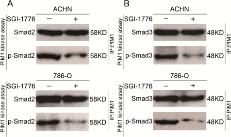
SGI-1776 purchased from MedChemExpress. Usage Cited in: Cell Death Dis. 2018 Feb 22;9(3):307. [Abstract]
PIM1 directly phosphorylated Smad2 and Smad3 and that inhibition of PIM1 kinase activity with SGI-1776 significantly reduces the protein levels of p-Smad2 and p-Smad3
-
Phytomedicine
6''-O-acetylsaikosaponin D targets STAT3-mediated transcriptional remodeling to induce ferroptosis and apoptosis. [Abstract]2026 May 15:157:158301. PMID: 42166974 -
Cell Chem Biol
2024 Feb 15;31(2):326-337.e11. PMID: 38016478 -
J Med Chem
Discovery of a Dual Tubulin and Poly(ADP-Ribose) Polymerase-1 Inhibitor by Structure-Based Pharmacophore Modeling, Virtual Screening, Molecular Docking, and Biological Evaluation. [Abstract]2021 Nov 11;64(21):15702-15715. PMID: 34670362 -
Front Cell Infect Microbiol
ZIKV prM hijacks PIM1 kinase for phosphorylation to prevent ubiquitin-mediated degradation and facilitate viral replication. [Abstract]2024 Nov 29:14:1502770. PMID: 39679197 -
-
eNeuro
2019 Aug 22;6(4):ENEURO.0003-19.2019. PMID: 31387876 -
Anticancer Drugs
2024 Jan 1;35(1):46-54. PMID: 37449977 -


Purity & Documentation
References
[1]. Park YK, et al. The novel anti-adipogenic effect and mechanisms of action of SGI-1776, a Pim-specific inhibitor, in 3T3-L1 adipocytes. International Journal of Molecular Medicine. 2016 Jan 1;37(1):157-64. [Content Brief]
[2]. Chen LS, et al. Mechanisms of cytotoxicity to Pim kinase inhibitor, SGI-1776, in acute myeloid leukemia. Blood. 2011 Jul 21;118(3):693-702. [Content Brief]
[3]. Hou X, et al. Biochemical changes of salivary gland adenoid cystic carcinoma cells induced by SGI-1776. Experimental cell research. 2017 Mar 15;352(2):403-411. [Content Brief]

Calculators
Concentration (start) × Volume (start) = Concentration (final) × Volume (final)


SGI-1776 Related Classifications
HY-13287A Related Classifications
 Powered by Bioz