



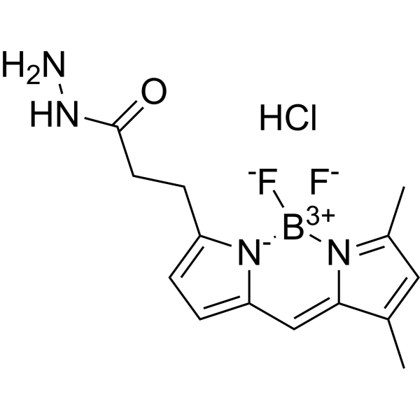
BODIPY FL hydrazide hydrochloride
BODIPY FL hydrazide hydrochloride is a BODIPY-containing hydrazide fluorescent dye. BODIPY FL hydrazide hydrochloride is used to label carbonyl groups on oxidatively damaged proteins, 3-ketosteroids, and heparin/heparan sulfate carbohydrates. BODIPY FL hydrazide hydrochloride specifically binds to protein carbonyls, steroid keto groups, or reducing-end carbonyls of carbohydrates through derivatization or reductive amination via its hydrazide group to form stable conjugates; after the reaction is complete, the labeled molecules emit strong fluorescence (Ex/Em = 488-495/516-520 nm).
For research use only. We do not sell to patients.
- CAS No.: 2183473-45-0
- Formula: C14H18BClF2N4O
- Molecular Weight:342.58
-
Storage:
Please store the product under the recommended conditions in the Certificate of Analysis.

To place orders, for customer services and technical support, please contact: MedChemExpress USA
Tel: 609-228-6898 E-mail: [email protected] [email protected]


-
 Biological Activity
-
Chemical Information
- Purity & Documentation
- References
-
 Help & FAQs


Biological Activity
Description
In Vitro
1. Stock solution preparation
1.1 Solvent: Dissolve BODIPY FL hydrazide hydrochloride in phosphate buffer (pH 7.5)[1], ethanol[2], or methanol[3], depending on the downstream application.
1.2 Concentration recommendations: Store at a high concentration of 1-10 mM in phosphate buffer (pH 7.5)[1]. Prepare stock solutions at 1 mg/mL in ethanol (dissolve 0.5 mg reagent in 5.0 mL ethanol[2]; use within 3 h), or at 1.25 mg/mL in methanol (store at −20°C)[3].
2. Working solution preparation
2.1 Diluent: For protein carbonyl labeling, dilute the stock solution with 125 mM phosphate buffer (pH 7.5)[1]; for steroid derivatization, use ethanol[2]; for glycan labeling, add the methanol stock solution directly to lyophilized disaccharide samples (subsequently remove methanol by vacuum centrifugation)[3].
2.2 Working concentration: For protein carbonyl labeling, use at a final concentration of 0.17 mM in the reaction mixture[1]; for steroid derivatization, use 100 µg/mL (molar ratio of 200 relative to progesterone)[2]; for glycan labeling, use 10 µL per 10 nmol disaccharide[3].
2.3 Note: Adjust the working solution concentration as needed; prepare fresh before use.
3. Staining procedure
3.1 For biological fluid samples (temporomandibular joint lavage fluid and diluted serum)[1]:
3.1.1 Use a 60 μL aliquot of temporomandibular joint lavage fluid or diluted serum per reaction.
3.1.2 Incubate with 0.17 mM BODIPY FL hydrazide hydrochloride in the presence of 125 mM phosphate buffer (pH 7.5) in a total reaction volume of 77 μL at 37°C for 2 h.
3.1.3 Perform the reaction and subsequent electrophoresis under light-protected conditions.
3.1.4 Subject the reaction mixture to SDS-PAGE after incubation.
3.2 For serum steroid samples[2]:
3.2.1 Add 10 µL steroid solution (final concentration 100 ng/mL) to 1 mL serum, dilute to 3 mL with water, add 10 mL dichloromethane, mix for 1 min, and let stand for 10 min.
3.2.2 Collect 8 mL of the dichloromethane layer and evaporate to dryness.
3.2.3 Add 0.2 mL BODIPY FL hydrazide hydrochloride solution and 0.2 mL 0.08% trifluoroacetic acid solution, and let stand at room temperature (approximately 22°C) for 15 h.
3.2.4 Inject a 10 µL volume into the HPLC instrument.
3.3 For lyophilized unsaturated heparin/heparan sulfate disaccharides[3]:
3.3.1 Prepare disaccharides by enzymatic digestion: add 2.5 mU (10 μl aliquot) each of heparinase I, II, and III per mg sample, add 40 μl 5× lyase buffer, bring to 1× with HPLC water in a total volume of 200 μl, incubate at 36°C for 12 h, and terminate the reaction by heating at 100°C for 5 min.
3.3.2 Equilibrate an Amersham PD-10 column with 30 mL HPLC water, load the sample, add 1.8 mL HPLC water and allow the effluent to flow to waste, add 3.5 mL HPLC water and collect the effluent, then lyophilize.
3.3.3 Add 10 μl (per 10 nmol disaccharide) of BODIPY FL hydrazide hydrochloride in methanol to the lyophilized sample, and evaporate the methanol by vacuum centrifugation at 36°C for 30 min.
3.3.4 Add 5 μl labeling solution (dimethyl sulfoxide:glacial acetic acid, 17:3, v/v) to the disaccharide/fluorophore mixture, and incubate at room temperature for 4 h.
3.3.5 Add 5 μl reducing agent (1 M NaBH4), and incubate at room temperature for 30 min.
3.3.6 Snap-freeze with liquid nitrogen, lyophilize to dryness, and resuspend in 8 μl reconstitution solution (DMSO:water, 1:1, v/v) before analysis.
4. Controls
4.1 Include diluted serum samples as a comparison control to assess the extent of carbonyl labeling relative to lavage samples[1].
5. Detection and analysis
5.1 Instrument type: For protein carbonyl labeling, use a UV light box with gel photography and image analysis software[1]; for steroid derivatization, use HPLC with fluorescence detection[2]; for disaccharide analysis, use a gradient HPLC system with an online fluorescence detector[3].
5.2 Excitation/emission wavelengths: 495 nm/516 nm for steroid derivatives[2]; 488 nm/520 nm for labeled disaccharides[3].
5.3 Result analysis: For protein carbonyls, quantify the extent of fluorescent labeling and Coomassie staining, and compare the extent of carbonyl labeling of each protein band between lavage fluid and serum samples; compare optical density units only within the same experiment (same polyacrylamide gel), as units depend on staining/destaining and fluorescence quenching; expect labeling of a 70-kd protein band (presumed to be albumin) and high-molecular-weight bands in the 100 to 200-kd range; the extent of carbonyl labeling per albumin band is higher in lavage fluid samples than in serum samples; note the rapid photobleaching of high-molecular-weight bandssup>[1]. For steroid derivatives, the BODIPY FL hydrazide hydrochloride derivative of progesterone is detected in acetonitrile-water (7 + 3) at 495 nm/516 nm; the detection limits are 550 fmoL for 17-hydroxyprogesterone, 550 fmoL for progesterone, 1.2 pmoL for dehydroepiandrosterone, 1.2 pmol for testosterone, and 3.7 pmoL for androstenedione (10 µL injection, signal-to-noise ratio = 5); the calibration range extends up to 1000 ng/mL[2]. For disaccharides, all eight fluorescently labeled Δ-disaccharides are separated and detected using a linear sodium chloride gradient on an isocratic sodium hydroxide mobile phase; comparison of sample digests with derivatized commercially available known disaccharide standards provides reference chromatograms of elution times; derivatization proceeds via reductive amination, involving the reducing-end carbonyl of the disaccharide and the hydrazide amine group of the fluorophore[3].
MedChemExpress (MCE) has not independently confirmed the accuracy of these methods. They are for reference only.


Chemical Information
-
CAS No. 2183473-45-0
-
Molecular Weight 342.58
-
Formula C14H18BClF2N4O
-
SMILES
O=C(CCC1=CC=C2C=C3C(C)=CC(C)=[N]3[B+3]([F-])([N-]21)[F-])NN.Cl
-
Shipping
Room temperature in continental US; may vary elsewhere.
-
Storage
Please store the product under the recommended conditions in the Certificate of Analysis.


Purity & Documentation
References
[2]. Katayama M, et al. Determination of progesterone and 17-hydroxyprogesterone by high performance liquid chromatography after pre-column derivatization with 4,4-difluoro-5,7-dimethyl-4-bora-3a,4a-diaza-s-indacene-3-propionohydra zide. The Analyst. 1998 Nov;123(11):2339-42. [Content Brief]
[3]. Skidmore M, et al. Labelling heparan sulphate saccharides with chromophore, fluorescence and mass tags for HPLC and MS separations. Methods in molecular biology (Clifton, N.J.). 2009;534:157-69. [Content Brief]

Calculators
Concentration (start) × Volume (start) = Concentration (final) × Volume (final)


BODIPY FL hydrazide hydrochloride Related Classifications
HY-D1332 Related Classifications
 Powered by Bioz